Department of Infection, Immunity and Inflammation, Institute for Lung Health, University of Leicester, Leicester, United Kingdom.
J Allergy Clin Immunol. 2011 Dec;128(6):1303-1309.e2. doi: 10.1016/j.jaci.2011.07.047. Epub 2011 Aug 27.
Fibrocytes are bone marrow-derived CD34(+) collagen I-positive cells present in peripheral blood that develop α-smooth muscle actin expression and contractile activity in tissue culture. They are implicated in the pathogenesis of tissue remodeling and fibrosis in both patients with asthma and those with idiopathic pulmonary fibrosis. Targeting fibrocyte migration might therefore offer a new approach for the treatment of these diseases. Ion channels play key roles in cell function, but the ion-channel repertoire of human fibrocytes is unknown.
We sought to examine whether human fibrocytes express the K(Ca)3.1 K(+) channel and to determine its role in cell differentiation, survival, and migration.
Fibrocytes were cultured from the peripheral blood of healthy subjects and patients with asthma. Whole-cell patch-clamp electrophysiology was used for the measurement of ion currents, whereas mRNA and protein were examined to confirm channel expression. Fibrocyte migration and proliferation assays were performed in the presence of K(Ca)3.1 ion-channel blockers.
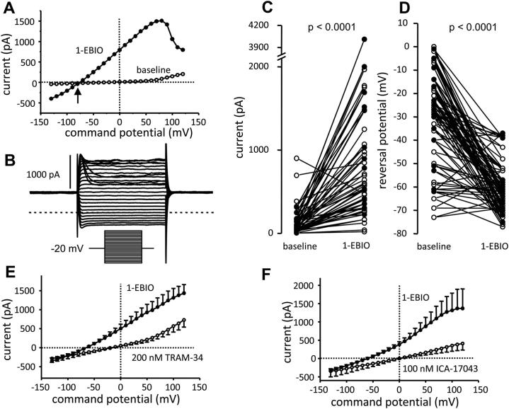
Human fibrocytes cultured from the peripheral blood of both healthy control subjects and asthmatic patients expressed robust K(Ca)3.1 ion currents together with K(Ca)3.1 mRNA and protein. Two specific and distinct K(Ca)3.1 blockers (TRAM-34 and ICA-17043) markedly inhibited fibrocyte migration in transwell migration assays. Channel blockers had no effect on fibrocyte growth, apoptosis, or differentiation in cell culture.
The K(+) channel K(Ca)3.1 plays a key role in human fibrocyte migration. Currently available K(Ca)3.1-channel blockers might therefore attenuate tissue fibrosis and remodeling in patients with diseases such as idiopathic pulmonary fibrosis and asthma through the inhibition of fibrocyte recruitment.
纤维细胞是骨髓来源的 CD34(+)胶原蛋白 I 阳性细胞,存在于外周血中,在组织培养中会发展出 α-平滑肌肌动蛋白表达和收缩活性。它们与哮喘患者和特发性肺纤维化患者的组织重塑和纤维化的发病机制有关。因此,靶向纤维细胞迁移可能为这些疾病的治疗提供新方法。离子通道在细胞功能中发挥关键作用,但人类纤维细胞的离子通道谱尚不清楚。
我们试图研究人类纤维细胞是否表达 K(Ca)3.1 K(+)通道,并确定其在细胞分化、存活和迁移中的作用。
从健康受试者和哮喘患者的外周血中培养纤维细胞。使用全细胞膜片钳电生理学测量离子电流,同时检查 mRNA 和蛋白质以确认通道表达。在存在 K(Ca)3.1 离子通道阻滞剂的情况下进行纤维细胞迁移和增殖测定。
从健康对照受试者和哮喘患者的外周血中培养的人类纤维细胞表达了强大的 K(Ca)3.1 离子电流,以及 K(Ca)3.1 mRNA 和蛋白质。两种特异性和不同的 K(Ca)3.1 阻滞剂(TRAM-34 和 ICA-17043)在 Transwell 迁移测定中显著抑制纤维细胞迁移。通道阻滞剂对纤维细胞生长、凋亡或细胞培养中的分化没有影响。
K(+)通道 K(Ca)3.1 在人类纤维细胞迁移中起关键作用。目前可用的 K(Ca)3.1 通道阻滞剂可能通过抑制纤维细胞募集来减轻特发性肺纤维化和哮喘等疾病患者的组织纤维化和重塑。