1. Russell H. Morgan Department of Radiology and Radiological Science, Division of MR Research, Johns Hopkins University School of Medicine, Baltimore, Maryland, United States.
1. Russell H. Morgan Department of Radiology and Radiological Science, Division of MR Research, Johns Hopkins University School of Medicine, Baltimore, Maryland, United States. ; 2. Cellular Imaging Section and Vascular Biology Program, Institute for Cell Engineering, Johns Hopkins University School of Medicine, Baltimore, Maryland, United States.
Theranostics. 2013 Dec 17;3(11):916-26. doi: 10.7150/thno.6943. eCollection 2013.
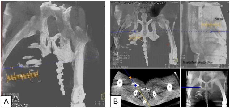
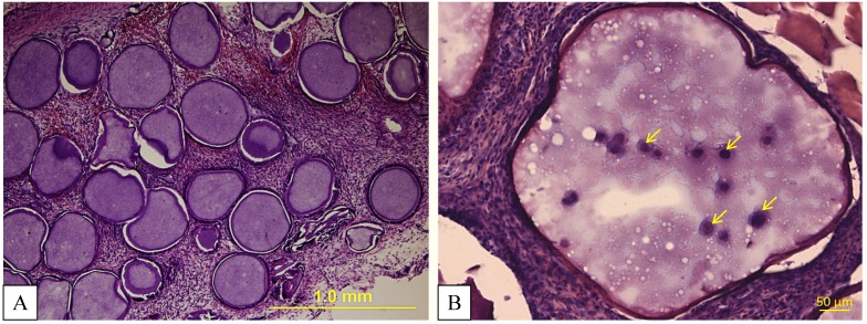
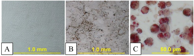
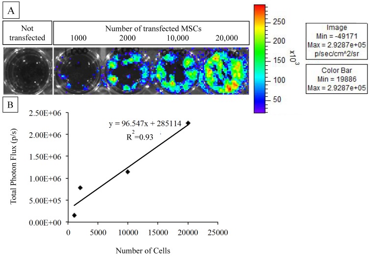
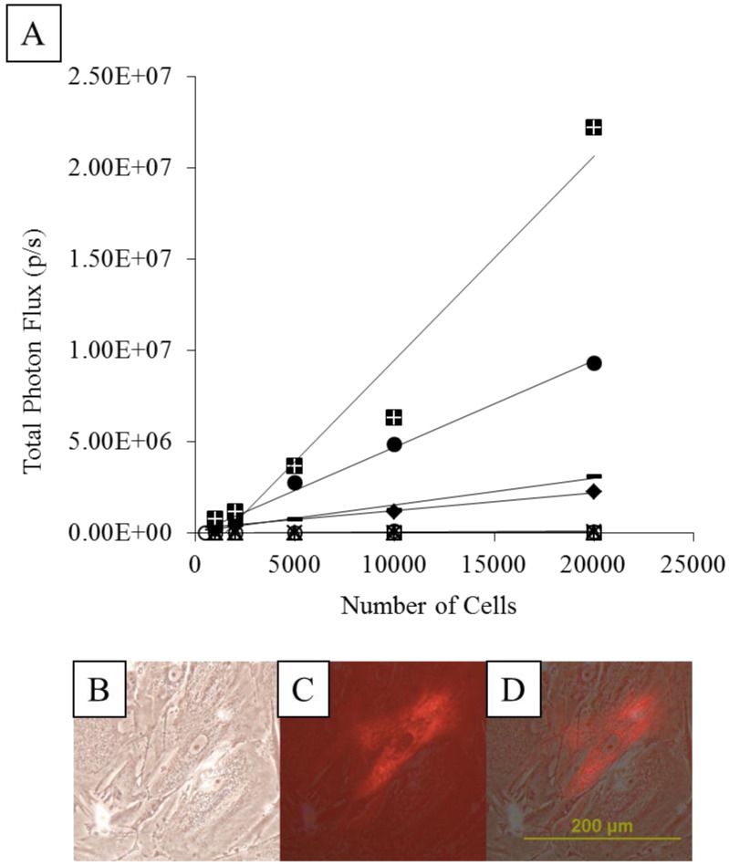
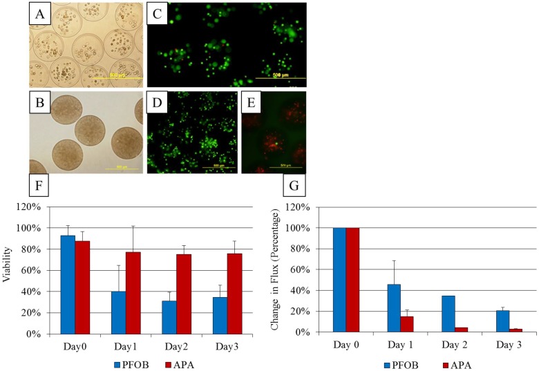
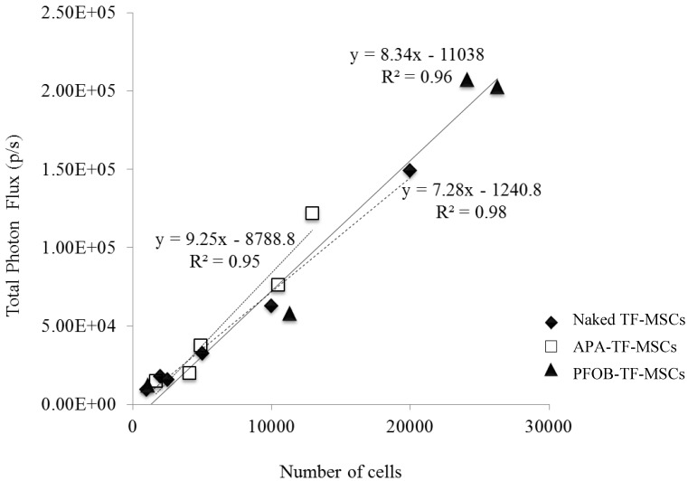
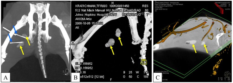
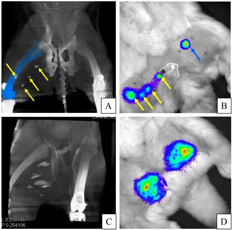
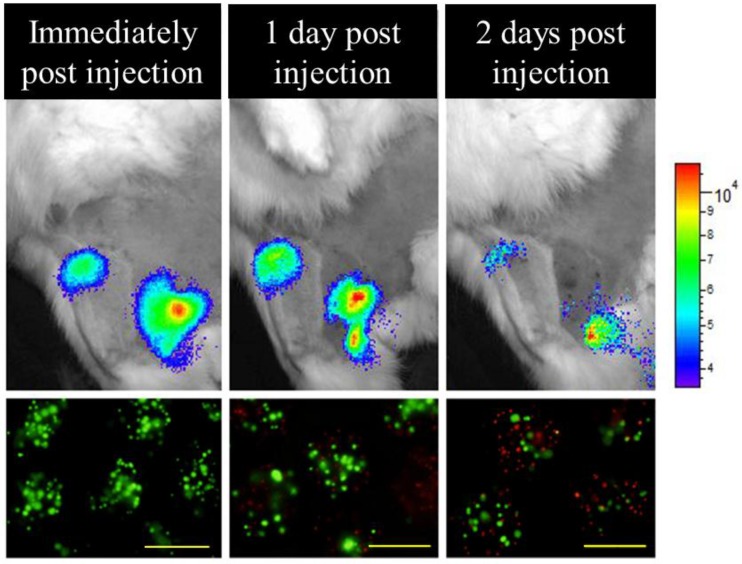
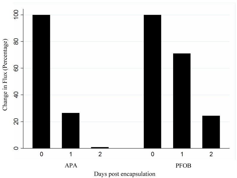
Poor cell survival and difficulties with visualization of cell delivery are major problems with current cell transplantation methods. To protect cells from early destruction, microencapsulation methods have been developed. The addition of a contrast agent to the microcapsule also could enable tracking by MR, ultrasound, and X-ray imaging. However, determining the cell viability within the microcapsule still remains an issue. Reporter gene imaging provides a way to determine cell viability, but delivery of the reporter probe by systemic injection may be hindered in ischemic diseases. In the present study, mesenchymal stem cells (MSCs) were transfected with triple fusion reporter gene containing red fluorescent protein, truncated thymidine kinase (SPECT/PET reporter) and firefly luciferase (bioluminescence reporter). Transfected cells were microencapsulated in either unlabeled or perfluorooctylbromide (PFOB) impregnated alginate. The addition of PFOB provided radiopacity to enable visualization of the microcapsules by X-ray imaging. Before intramuscular transplantation in rabbit thigh muscle, the microcapsules were incubated with D-luciferin, and bioluminescence imaging (BLI) was performed immediately. Twenty-four and forty-eight hours post transplantation, c-arm CT was used to target the luciferin to the X-ray-visible microcapsules for BLI cell viability assessment, rather than systemic reporter probe injections. Not only was the bioluminescent signal emission from the PFOB-encapsulated MSCs confirmed as compared to non-encapsulated, naked MSCs, but over 90% of injection sites of PFOB-encapsulated MSCs were visible on c-arm CT. The latter aided in successful targeting of the reporter probe to injection sites using conventional X-ray imaging to determine cell viability at 1-2 days post transplantation. Blind luciferin injections to the approximate location of unlabeled microcapsules resulted in successful BLI signal detection in only 18% of injections. In conclusion, reporter gene probes can be more precisely targeted using c-arm CT for in vivo transplant viability assessment, thereby avoiding large and costly systemic injections of a reporter probe.
细胞存活率低和细胞递送可视化困难是当前细胞移植方法面临的主要问题。为了保护细胞免受早期破坏,已经开发出微囊化方法。向微胶囊中添加造影剂也可以使磁共振、超声和 X 射线成像能够进行跟踪。然而,确定微胶囊内的细胞活力仍然是一个问题。报告基因成像提供了一种确定细胞活力的方法,但是通过全身注射递送报告探针可能会在缺血性疾病中受到阻碍。在本研究中,间充质干细胞(MSCs)被转染了含有红色荧光蛋白、截断胸苷激酶(SPECT/PET 报告基因)和萤火虫荧光素酶(生物发光报告基因)的三重融合报告基因。转染的细胞被包裹在未标记或全氟辛基溴(PFOB)浸渍的藻酸盐中。添加 PFOB 可提供放射密度,使 X 射线成像能够可视化微胶囊。在兔大腿肌肉内移植前,将微胶囊与 D-荧光素孵育,并立即进行生物发光成像(BLI)。移植后 24 小时和 48 小时,使用 c 臂 CT 将荧光素靶向到 X 射线可见的微胶囊进行 BLI 细胞活力评估,而不是全身报告探针注射。与未包裹的裸 MSCs 相比,不仅证实了 PFOB 包裹的 MSCs 的生物发光信号发射,而且超过 90%的 PFOB 包裹的 MSCs 注射部位在 c 臂 CT 上可见。后者通过常规 X 射线成像辅助成功靶向报告探针到注射部位,以确定移植后 1-2 天的细胞活力。向未标记的微胶囊的大致位置盲目注射荧光素,仅 18%的注射能够成功检测到 BLI 信号。总之,使用 c 臂 CT 可以更精确地靶向报告基因探针,用于体内移植活力评估,从而避免了昂贵的全身注射报告探针。