1 Department of Radiology, Optical Molecular Imaging, Erasmus Medical Center, Rotterdam, the Netherlands.
2 Percuros BV, Enschede, the Netherlands.
Cell Transplant. 2017 Dec;26(12):1878-1889. doi: 10.1177/0963689717739718.
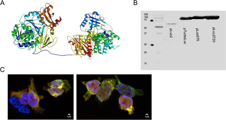
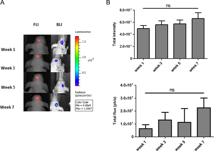
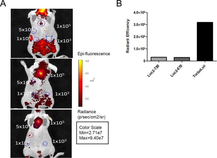
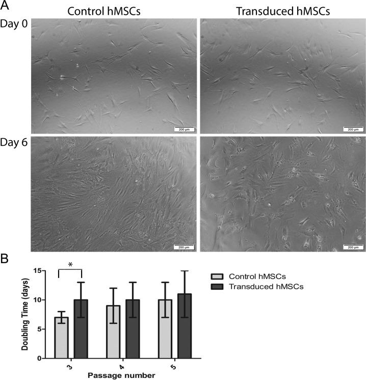
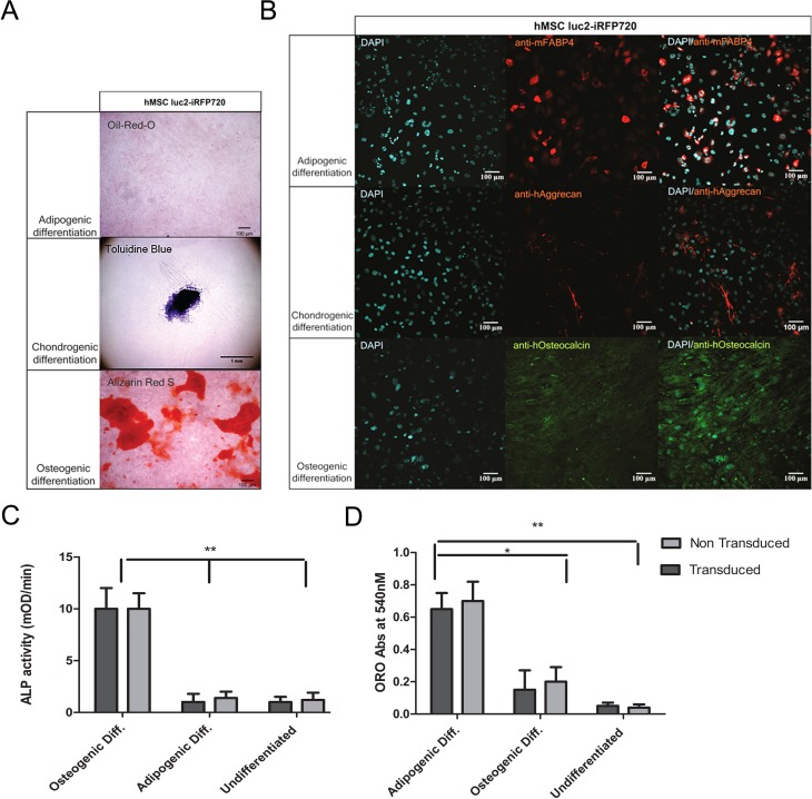
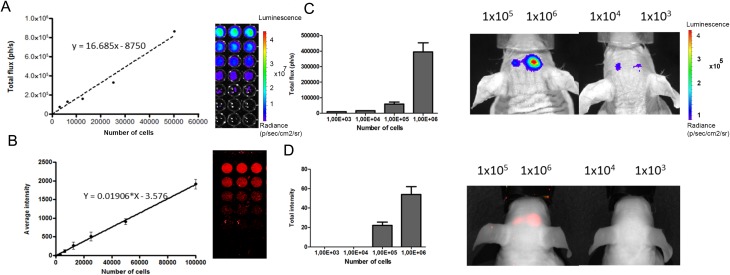
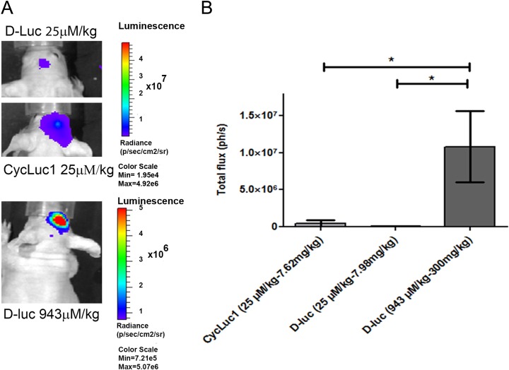
Biodistribution and fate of transplanted stem cells via longitudinal monitoring has been successfully achieved in the last decade using optical imaging. However, sensitive longitudinal imaging of transplanted stem cells in deep tissue like the brain remains challenging not only due to low light penetration but because of other factors such as low or inferior expression levels of optical reporters in stem cells and stem cell death after transplantation. Here we describe an optimized imaging protocol for sensitive long-term monitoring of bone marrow-derived human mesenchymal stem cells (hMSCs) expressing a novel bioluminescent/near infrared fluorescent (NIRF) fusion reporter transplanted in mouse brain cortex. Lentivirus expressing the luc2-iRFP720 reporter, a fusion between luc2 codon-optimized firefly luciferase (luc2) and the gene encoding NIRF protein iRFP720, was generated to transduce hMSCs. These cells were analyzed for their fluorescent and bioluminescent emission and checked for their differentiation potential. In vivo experiments were performed by transplanting decreasing amounts of luc2-iRFP720 expressing hMSCs in mouse brain, followed by fluorescence and bioluminescence imaging (BLI) starting 1 wk after cell injection when the blood-brain barrier was restored. Bioluminescent images were acquired when signals peaked and used to compare different luc2 substrate performances, that is, D-luciferin (D-Luc; 25 μM/kg or 943 μM/kg) or CycLuc1 (25 μM/kg). Results showed that luc2-iRFP720 expressing hMSCs maintained a good in vitro differentiation potential toward adipocytes, chondrocytes, and osteocytes, suggesting that lentiviral transduction did not affect cell behavior. Moreover, in vivo experiments allowed us to image as low as 1 × 10 cells using both fluorescence and BLI. The highest bioluminescent signals (∼1 × 10 photons per second) were achieved 15 min after the injection of D-Luc (943 μM/kg). This allowed us to monitor as low as 1 × 10 hMSCs for the subsequent 7 wk without a significant drop in bioluminescent signals, suggesting the sustained viability of hMSCs transplanted into the cortex.
在过去的十年中,通过光学成像成功地实现了通过纵向监测移植干细胞的生物分布和命运。然而,由于光穿透率低以及其他因素(例如干细胞中光学报告基因的低表达或表达水平低,以及移植后干细胞死亡),在深部组织(如大脑)中对移植干细胞进行敏感的纵向成像仍然具有挑战性。在这里,我们描述了一种优化的成像方案,用于敏感地长期监测在小鼠大脑皮层中移植的表达新型生物发光/近红外荧光(NIRF)融合报告基因的骨髓源性人间充质干细胞(hMSC)。表达 luc2-iRFP720 报告基因的慢病毒(luc2 编码的优化萤火虫荧光素酶(luc2)与编码 NIRF 蛋白 iRFP720 的基因的融合)被生成以转导 hMSC。分析了这些细胞的荧光和生物发光发射,并检查了它们的分化潜力。通过在细胞注射后 1 周当血脑屏障恢复时,将表达 luc2-iRFP720 的逐渐减少量的 hMSC 移植到小鼠大脑中,然后进行荧光和生物发光成像(BLI),开始进行体内实验。当信号达到峰值时,获取生物发光图像,并用于比较不同 luc2 底物的性能,即 D-荧光素(D-Luc;25 μM/kg 或 943 μM/kg)或 CycLuc1(25 μM/kg)。结果表明,表达 luc2-iRFP720 的 hMSC 维持了向脂肪细胞,软骨细胞和成骨细胞的良好体外分化潜力,表明慢病毒转导不会影响细胞行为。此外,体内实验使我们能够使用荧光和 BLI 成像低至 1×10 个细胞。在注射 D-Luc(943 μM/kg)后 15 分钟,获得了最高的生物发光信号(约 1×10 个光子/秒)。这使我们能够在没有生物发光信号明显下降的情况下监测随后的 7 周内低至 1×10 hMSC,表明移植到皮层中的 hMSC 具有持续的活力。