Department of Neurology, Zhengzhou People's Hospital, Zhengzhou 450003, China.
BMC Neurol. 2014 Jan 16;14:14. doi: 10.1186/1471-2377-14-14.
Tortuous blood vessels are commonly seen in the cerebral arteries. The association between vertebrobasilar artery tortuosity and vascular vertigo remains obscure.
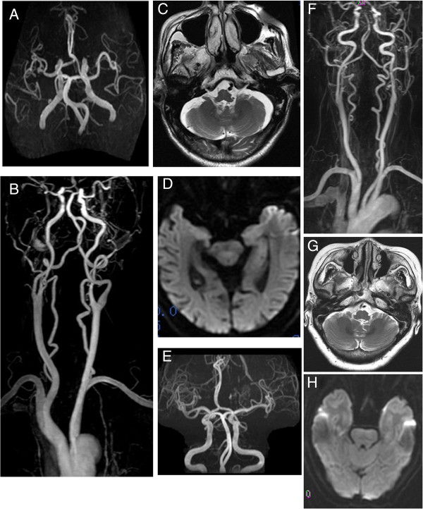
We describe two patients with vascular vertigo who had bilateral curving and spiral looping in multiple segments of the vertebral arteries and also exhibited basilar artery tortuosity. Both patients had cerebrovascular risk factors and exhibited clinical features of vertigo with high severity, slow recovery, and recurrent tendencies. Contrast enhanced magnetic resonance angiography of the neck showed bilateral tortuosity in the V2 segments and spiral twisting in the V4 segments of the vertebral arteries, and basilar artery curving. No obvious sign of atherosclerotic stenosis was found in the vertebrobasilar arteries and no abnormalities were observed in the internal carotid arteries. Transcranial Doppler ultrasound showed decreased blood flow in tortuous vertebrobasilar arteries. Brainstem auditory evoked potentials showed that the interpeak latencies (IPL) of waves III-IV were prolonged, with a ratio of IPL III-V/IPL I-III > 1.
Vertebrobasilar tortuosity in combination with cerebrovascular risk factors may lead to vascular vertigo in these patients.
脑动脉中常可见迂曲的血管。椎基底动脉迂曲与血管性眩晕之间的关系尚不清楚。
我们描述了 2 例血管性眩晕患者,他们的椎动脉在多个节段出现双侧弯曲和螺旋状弯曲,基底动脉也出现迂曲。这 2 例患者均有脑血管危险因素,表现为眩晕的临床特征为严重度高、恢复缓慢、反复发作。颈部增强磁共振血管造影显示双侧椎动脉 V2 段迂曲,V4 段螺旋扭曲,基底动脉弯曲。椎基底动脉无明显粥样硬化狭窄征象,颈内动脉无异常。经颅多普勒超声显示迂曲的椎基底动脉血流减少。脑干听觉诱发电位显示波 III-IV 的峰间潜伏期(IPL)延长,IPL III-V/IPL I-III 比值>1。
椎基底动脉迂曲合并脑血管危险因素可能导致这些患者出现血管性眩晕。