Department of Veterinary Medicine, College of Veterinary Medicine, National Chung-Hsing University, No, 250, Kuo Kuang Road, Taichung 402, Taiwan.
BMC Neurosci. 2014 Jan 17;15:15. doi: 10.1186/1471-2202-15-15.
Hepatic encephalopathy (HE) is a reversible neuropsychiatric syndrome associated with acute and chronic liver diseases. It includes a number of neuropsychiatric disturbances including impaired motor activity and coordination, intellectual and cognitive function.
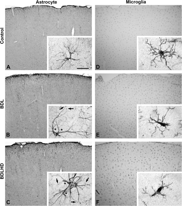
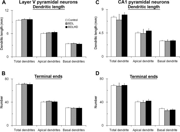
In the present study, we used a chronic rat HE model by ligation of the bile duct (BDL) for 4 weeks. These rats showed increased plasma ammonia level, bile duct hyperplasia and impaired spatial learning memory and motor coordination when tested with Rota-rod and Morris water maze tests, respectively. By immunohistochemistry, the cerebral cortex showed swelling of astrocytes and microglia activation. To gain a better understanding of the effect of HE on the brain, the dendritic arbors of layer V cortical pyramidal neurons and hippocampal CA1 pyramidal neurons were revealed by an intracellular dye injection combined with a 3-dimensional reconstruction. Although the dendritic arbors remained unaltered, the dendritic spine density on these neurons was significantly reduced. It was suggested that the reduction of dendritic spines may be the underlying cause for increased motor evoked potential threshold and prolonged central motor conduction time in clinical finding in cirrhosis.
We found that HE perturbs CNS functions by altering the dendritic morphology of cortical and hippocampal pyramidal neurons, which may be the underlying cause for the motor and intellectual impairments associated with HE patients.
肝性脑病(HE)是一种与急性和慢性肝脏疾病相关的可逆转的神经精神综合征。它包括多种神经精神障碍,包括运动活动和协调受损、智力和认知功能受损。
在本研究中,我们使用胆管结扎(BDL)4 周的慢性大鼠 HE 模型。这些大鼠表现为血浆氨水平升高、胆管增生以及在旋转棒和 Morris 水迷宫测试中分别出现空间学习记忆和运动协调受损。通过免疫组织化学,大脑皮层显示星形胶质细胞肿胀和小胶质细胞激活。为了更好地了解 HE 对大脑的影响,我们通过细胞内染料注射结合三维重建来揭示 V 层皮质锥体神经元和海马 CA1 锥体神经元的树突树。尽管树突树保持不变,但这些神经元上的树突棘密度显著降低。这表明,在肝硬化患者中运动诱发电位阈值增加和中枢运动传导时间延长的临床发现中,树突棘的减少可能是潜在的原因。
我们发现 HE 通过改变皮质和海马锥体神经元的树突形态扰乱中枢神经系统功能,这可能是与 HE 患者相关的运动和智力障碍的潜在原因。