Sinha Sanjeev, Ekka Meera, Sharma Uma, P Raghunandan, Pandey R M, Jagannathan N R
Department of Medicine, All India Institute of Medical Sciences, Ansari Nagar, New Delhi 110029, India.
BMC Res Notes. 2014 Jan 17;7:41. doi: 10.1186/1756-0500-7-41.
The brain is a target for diabetic end-organ damage, though the pathophysiology of diabetic encephalopathy is still not well understood. The aim of the present study was to investigate the effect of diabetes on the metabolic profile of brain of patients having diabetes in comparison to healthy controls, using in-vivo magnetic resonance spectroscopy to get an insight into the pathophysiology of cerebral damages caused due to diabetes.
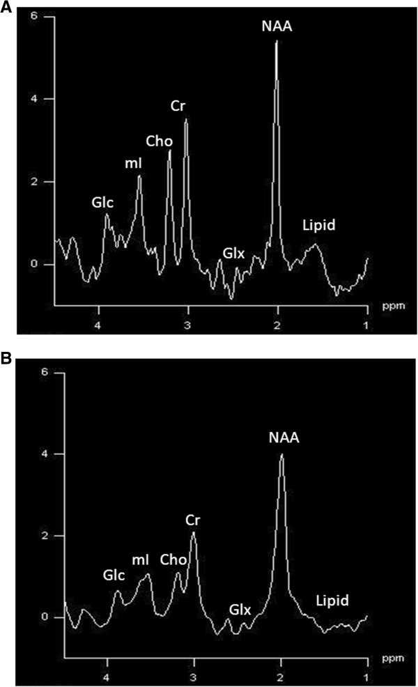
Single voxel proton magnetic resonance spectroscopy (1H-MRS) was performed at 1.5 T on right frontal, right parieto-temporal and right parieto-occipital white matter regions of the brain of 10 patients having type-2 diabetes along with 7 healthy controls. Absolute concentration of N-acetylaspartate (NAA), choline (cho), myo-inositol (mI), glutamate (Glu) and glutamine (Gln), creatine (Cr) and glucose were determined using the LC-Model and compared between the two groups.
The concentration of N-acetylaspartate was significantly lower in the right frontal [4.35 ±0.69 vs. 5.23 ±0.74; p = 0.03] and right parieto-occipital region [5.44 ±0.52 vs.6.08 ±0.25; p = 0.02] of the brain of diabetics as compared to the control group. The concentrations of glutamate and glutamine were found to be significantly higher in the right frontal region of the brain [7.98 ±2.57 vs. 5.32 ±1.43; P = 0.01] in diabetics. Glucose levels were found significantly elevated in all the three regions of the brain in diabetics as compared to the control group. However, no significant changes in levels of choline, myo-inositol and creatine were observed in the three regions of the brain examined among the two groups.
1H-MRS analysis indicates that type-2 diabetes mellitus may cause subtle changes in the metabolic profile of the brain. Decreased concentrations of NAA might be indicative of decreased neuronal viability in diabetics while elevated concentrations of Gln and Glu might be related to the fluid imbalance resulting from disruption of glucose homeostasis.
大脑是糖尿病终末器官损伤的靶器官,尽管糖尿病性脑病的病理生理学仍未完全明确。本研究的目的是通过体内磁共振波谱技术,对比糖尿病患者与健康对照者,探究糖尿病对大脑代谢谱的影响,从而深入了解糖尿病所致脑损伤的病理生理学机制。
对10例2型糖尿病患者及7名健康对照者的大脑右侧额叶、右侧顶颞叶和右侧顶枕叶白质区域进行1.5T磁共振单体素质子磁共振波谱(1H-MRS)检查。使用LC-Model测定N-乙酰天门冬氨酸(NAA)、胆碱(Cho)、肌醇(mI)、谷氨酸(Glu)、谷氨酰胺(Gln)、肌酸(Cr)和葡萄糖的绝对浓度,并在两组间进行比较。
与对照组相比,糖尿病患者大脑右侧额叶[4.35±0.69 vs. 5.23±0.7;p = 0.03]和右侧顶枕叶区域[5.44±0.52 vs. 6.08±0.25;p = 0.02]的N-乙酰天门冬氨酸浓度显著降低。糖尿病患者大脑右侧额叶区域的谷氨酸和谷氨酰胺浓度显著升高[7.98±2.57 vs. 5.32±1.43;P = 0.01]。与对照组相比,糖尿病患者大脑所有三个区域的葡萄糖水平均显著升高。然而,两组间所检查的大脑三个区域的胆碱、肌醇和肌酸水平均未观察到显著变化。
1H-MRS分析表明,2型糖尿病可能导致大脑代谢谱的细微变化。NAA浓度降低可能表明糖尿病患者神经元活力下降,而Gln和Glu浓度升高可能与葡萄糖稳态破坏导致的液体失衡有关。