Rondón-Lagos Milena, Verdun Di Cantogno Ludovica, Marchiò Caterina, Rangel Nelson, Payan-Gomez Cesar, Gugliotta Patrizia, Botta Cristina, Bussolati Gianni, Ramírez-Clavijo Sandra R, Pasini Barbara, Sapino Anna
Department of Medical Sciences, University of Turin, Via Santena 7, 10126 Turin, Italy.
Mol Cytogenet. 2014 Jan 23;7(1):8. doi: 10.1186/1755-8166-7-8.
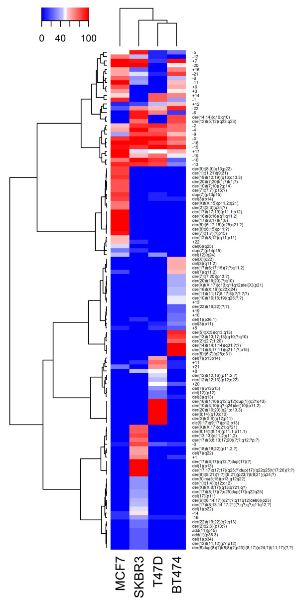
The MCF7 (ER+/HER2-), T47D (ER+/HER2-), BT474 (ER+/HER2+) and SKBR3 (ER-/HER2+) breast cancer cell lines are widely used in breast cancer research as paradigms of the luminal and HER2 phenotypes. Although they have been subjected to cytogenetic analysis, their chromosomal abnormalities have not been carefully characterized, and their differential cytogenetic profiles have not yet been established. In addition, techniques such as comparative genomic hybridization (CGH), microarray-based CGH and multiplex ligation-dependent probe amplification (MLPA) have described specific regions of gains, losses and amplifications of these cell lines; however, these techniques cannot detect balanced chromosomal rearrangements (e.g., translocations or inversions) or low frequency mosaicism.
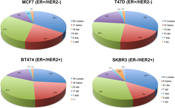
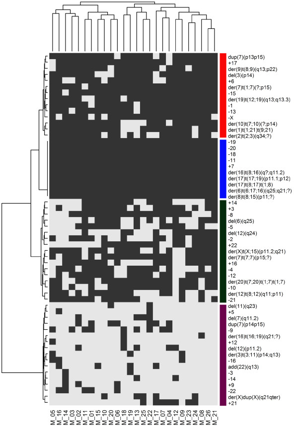
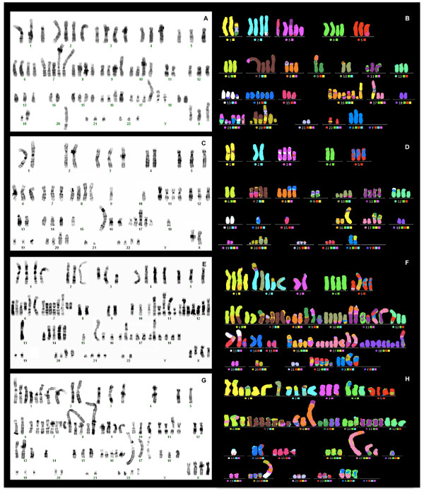
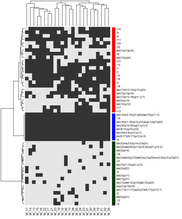
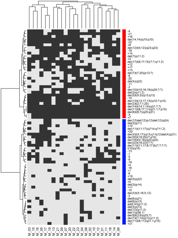
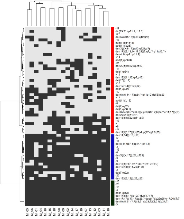
A range of 19 to 26 metaphases of the MCF7, T47D, BT474 and SKBR3 cell lines was studied using conventional (G-banding) and molecular cytogenetic techniques (multi-color fluorescence in situ hybridization, M-FISH). We detected previously unreported chromosomal changes and determined the content and frequency of chromosomal markers. MCF7 and T47D (ER+/HER2-) cells showed a less complex chromosomal make up, with more numerical than structural alterations, compared to BT474 and SKBR3 (HER2+) cells, which harbored the highest frequency of numerical and structural aberrations. Karyotype heterogeneity and clonality were determined by comparing all metaphases within and between the four cell lines by hierarchical clustering. The latter analysis identified five main clusters. One of these clusters was characterized by numerical chromosomal abnormalities common to all cell lines, and the other four clusters encompassed cell-specific chromosomal abnormalities. T47D and BT474 cells shared the most chromosomal abnormalities, some of which were shared with SKBR3 cells. MCF7 cells showed a chromosomal pattern that was markedly different from those of the other cell lines.
Our study provides a comprehensive and specific characterization of complex chromosomal aberrations of MCF7, T47D, BT474 and SKBR3 cell lines.The chromosomal pattern of ER+/HER2- cells is less complex than that of ER+/HER2+ and ER-/HER2+ cells. These chromosomal abnormalities could influence the biologic and pharmacologic response of cells. Finally, although gene expression profiling and aCGH studies have classified these four cell lines as luminal, our results suggest that they are heterogeneous at the cytogenetic level.
MCF7(雌激素受体阳性/人表皮生长因子受体2阴性)、T47D(雌激素受体阳性/人表皮生长因子受体2阴性)、BT474(雌激素受体阳性/人表皮生长因子受体2阳性)和SKBR3(雌激素受体阴性/人表皮生长因子受体2阳性)乳腺癌细胞系作为管腔型和人表皮生长因子受体2表型的范例,在乳腺癌研究中被广泛应用。尽管它们已经接受了细胞遗传学分析,但其染色体异常尚未得到仔细表征,其不同的细胞遗传学图谱也尚未建立。此外,诸如比较基因组杂交(CGH)、基于微阵列的CGH和多重连接依赖探针扩增(MLPA)等技术已经描述了这些细胞系的特定区域的增益、缺失和扩增情况;然而,这些技术无法检测到平衡的染色体重排(例如易位或倒位)或低频嵌合体。
使用传统(G显带)和分子细胞遗传学技术(多色荧光原位杂交,M-FISH)研究了MCF7、T47D、BT474和SKBR3细胞系的19至26个中期相。我们检测到了先前未报告的染色体变化,并确定了染色体标记物的含量和频率。与BT474和SKBR3(人表皮生长因子受体2阳性)细胞相比,MCF7和T47D(雌激素受体阳性/人表皮生长因子受体2阴性)细胞的染色体组成较不复杂,数值改变多于结构改变,而BT474和SKBR3细胞的数值和结构畸变频率最高。通过层次聚类比较四个细胞系内部和之间的所有中期相来确定核型异质性和克隆性。后一项分析确定了五个主要聚类。其中一个聚类的特征是所有细胞系共有的数值染色体异常,另外四个聚类包含细胞特异性染色体异常。T47D和BT474细胞共享的染色体异常最多,其中一些与SKBR3细胞共享。MCF7细胞显示出与其他细胞系明显不同的染色体模式。
我们的研究提供了MCF7、T47D、BT474和SKBR3细胞系复杂染色体畸变的全面而具体的表征。雌激素受体阳性/人表皮生长因子受体2阴性细胞的染色体模式比雌激素受体阳性/人表皮生长因子受体2阳性和雌激素受体阴性/人表皮生长因子受体2阳性细胞的染色体模式更不复杂。这些染色体异常可能影响细胞的生物学和药理学反应。最后,尽管基因表达谱分析和阵列比较基因组杂交研究将这四个细胞系归类为管腔型,但我们的结果表明它们在细胞遗传学水平上是异质的。