Schepens Eye Research Institute/Massachusetts Eye and Ear and Department of Ophthalmology, Harvard Medical School, Boston, Massachusetts, United States of America.
Department of Ophthalmology, University of Pittsburgh School of Medicine, Pittsburgh, Pennsylvania, United States of America.
PLoS One. 2014 Jan 21;9(1):e86260. doi: 10.1371/journal.pone.0086260. eCollection 2014.
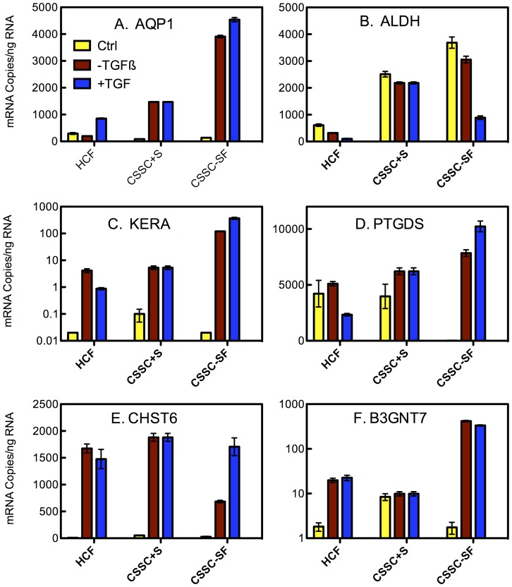
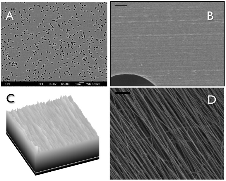
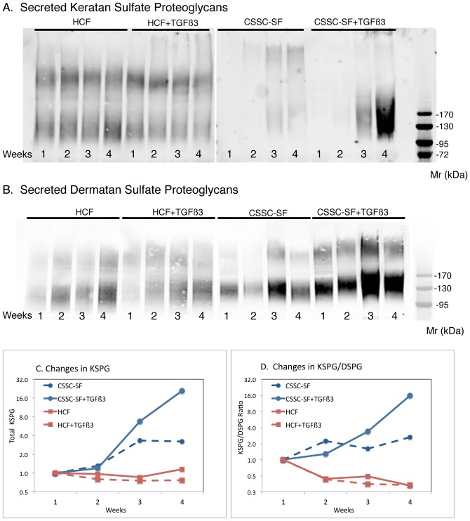
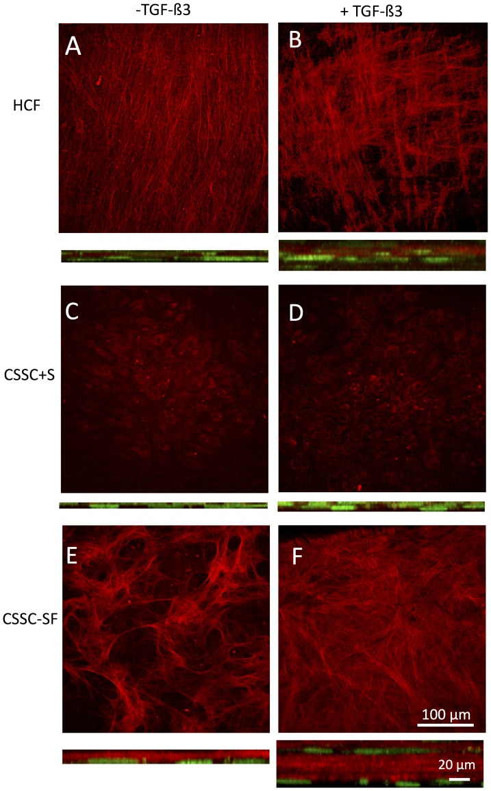
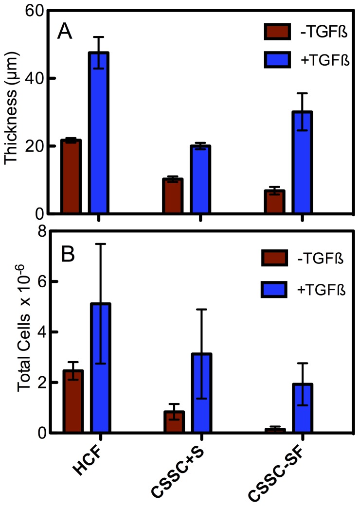
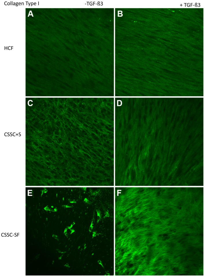
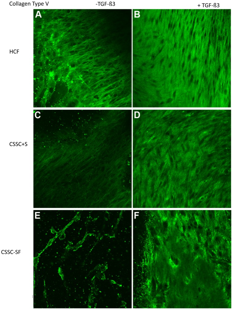
Human corneal fibroblasts (HCF) and corneal stromal stem cells (CSSC) each secrete and organize a thick stroma-like extracellular matrix in response to different substrata, but neither cell type organizes matrix on tissue-culture polystyrene. This study compared cell differentiation and extracellular matrix secreted by these two cell types when they were cultured on identical substrata, polycarbonate Transwell filters. After 4 weeks in culture, both cell types upregulated expression of genes marking differentiated keratocytes (KERA, CHST6, AQP1, B3GNT7). Absolute expression levels of these genes and secretion of keratan sulfate proteoglycans were significantly greater in CSSC than HCF. Both cultures produced extensive extracellular matrix of aligned collagen fibrils types I and V, exhibiting cornea-like lamellar structure. Unlike HCF, CSSC produced little matrix in the presence of serum. Construct thickness and collagen organization was enhanced by TGF-ß3. Scanning electron microscopic examination of the polycarbonate membrane revealed shallow parallel grooves with spacing of 200-300 nm, similar to the topography of aligned nanofiber substratum which we previously showed to induce matrix organization by CSSC. These results demonstrate that both corneal fibroblasts and stromal stem cells respond to a specific pattern of topographical cues by secreting highly organized extracellular matrix typical of corneal stroma. The data also suggest that the potential for matrix secretion and organization may not be directly related to the expression of molecular markers used to identify differentiated keratocytes.
人角膜成纤维细胞 (HCF) 和角膜基质干细胞 (CSSC) 分别在不同的基质上分泌和组织形成厚的基质样细胞外基质,但这两种细胞类型都不会在组织培养聚苯乙烯上组织基质。本研究比较了这两种细胞类型在相同基质(聚碳酸酯 Transwell 过滤器)上培养时的细胞分化和分泌的细胞外基质。培养 4 周后,两种细胞类型均上调了标记分化的角膜细胞 (KERA、CHST6、AQP1、B3GNT7) 的基因表达。CSSC 中这些基因的绝对表达水平和角膜硫酸乙酰肝素蛋白聚糖的分泌显著高于 HCF。两种培养物均产生大量排列整齐的 I 型和 V 型胶原纤维的细胞外基质,表现出类似角膜的层状结构。与 HCF 不同的是,CSSC 在存在血清的情况下产生的基质很少。TGF-ß3 增强了构建厚度和胶原组织的排列。聚碳酸酯膜的扫描电子显微镜检查显示出浅的平行凹槽,间距为 200-300nm,类似于我们之前展示的诱导 CSSC 基质组织的排列纳米纤维基底的形貌。这些结果表明,角膜成纤维细胞和基质干细胞都对特定的形貌线索模式做出反应,分泌具有典型角膜基质组织的高度有序的细胞外基质。数据还表明,基质分泌和组织的潜力可能与用于鉴定分化的角膜细胞的分子标志物的表达没有直接关系。