Flumerfelt B A, Kiernan J A, Krcek J P, Sholdice J
Department of Anatomy, University of Western Ontario, London, Canada.
J Anat. 1986 Jun;146:117-30.
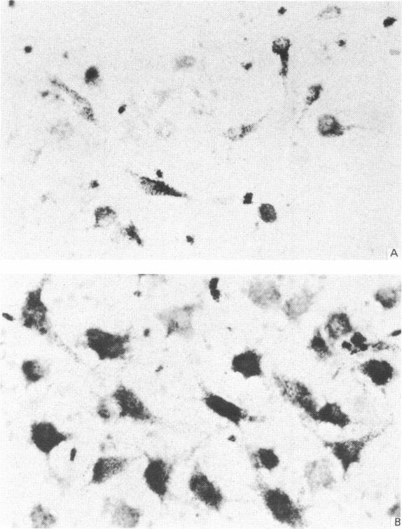
Reinnervation of the skeletal muscle in the tongue following vago-hypoglossal anastomosis was studied by means of retrograde labelling with horseradish peroxidase and anterograde labelling with the autoradiographic tracing method combined with acetylcholinesterase staining for motor endplates. The proximal stump of the transected vagus nerve was anastomosed to the distal stump of the transected hypoglossal nerve in the neck, or in the thorax below the emergence of the recurrent laryngeal fibres. After 2-3 months, reinnervation of the tongue by vagal fibres was studied. Control cases in which the hypoglossal nerve was transected, but anastomosis was not performed, revealed that innervation of the lingual muscle is derived entirely from the hypoglossal nerves. Following unilateral vago-hypoglossal anastomosis a reduced number of fine nerve fibres terminated in relation to the acetylcholinesterase-stained endplates on the side of the anastomosis. At no time were fibres on either side observed to form sprouts which crossed the midline. Horseradish peroxidase (HRP) was injected into the tongue to determine the origin of the fibres that reinnervated the lingual muscle following anastomosis. On the side of the anastomosis, HRP-labelled neurons were present within the dorsal motor nucleus of the vagus and were absent from the hypoglossal nucleus. When the anastomosis was performed in the neck, neurons within the nucleus ambiguous were also labelled with HRP, but this was not observed following anastomosis in the thorax below the recurrent laryngeal nerve. When tritiated amino acids were injected into the dorsal motor nucleus of the vagus, the motor endplates on the anastomosed side of the tongue were labelled autoradiographically. This labelling could not be abolished by transecting both hypoglossal nerves, confirming that the labelling was due to reinnervation by vagal fibres. It is concluded that anastomosis of the proximal end of the transected vagus nerve to the distal end of the transected hypoglossal nerve is followed by regeneration of the vagal fibres which cross the anastomosis and reinnervate the denervated motor endplates in the tongue. The cell bodies of origin are located within the dorsal motor nucleus of the vagus and are preganglionic parasympathetic neurons.
通过辣根过氧化物酶逆行标记以及将放射自显影示踪法与运动终板乙酰胆碱酯酶染色相结合的顺行标记方法,研究了迷走-舌下神经吻合术后舌部骨骼肌的再支配情况。将切断的迷走神经近端残端与颈部或喉返神经发出部位下方胸部的切断的舌下神经远端残端进行吻合。2至3个月后,研究迷走神经纤维对舌的再支配情况。切断舌下神经但未进行吻合的对照病例显示,舌肌的神经支配完全来自舌下神经。单侧迷走-舌下神经吻合术后,吻合侧乙酰胆碱酯酶染色终板处终止的细神经纤维数量减少。两侧均未观察到纤维形成穿过中线的新芽。将辣根过氧化物酶(HRP)注入舌内,以确定吻合术后重新支配舌肌的纤维来源。在吻合侧,HRP标记的神经元存在于迷走神经背运动核内,而舌下神经核内则没有。当在颈部进行吻合时,疑核内的神经元也被HRP标记,但在喉返神经下方胸部进行吻合后未观察到这种情况。当将氚标记的氨基酸注入迷走神经背运动核时,舌吻合侧的运动终板被放射自显影标记。切断双侧舌下神经并不能消除这种标记,证实这种标记是由于迷走神经纤维的再支配所致。得出的结论是,切断的迷走神经近端与切断的舌下神经远端吻合后,迷走神经纤维再生,穿过吻合口,重新支配舌内失神经的运动终板。其起源的细胞体位于迷走神经背运动核内,是节前副交感神经元。