Maekawa Kengo, Baba Tomoko, Otomo Sumi, Morishita Shoji, Tamura Nobushige
Department of Anesthesiology, Graduate School of Medical Sciences, Kumamoto University, Kumamoto, Japan ; Department of Anesthesiology, Kumamoto Chuo Hospital, Kumamoto, Japan.
Department of Anesthesiology, Kumamoto Chuo Hospital, Kumamoto, Japan.
PLoS One. 2014 Jan 27;9(1):e87375. doi: 10.1371/journal.pone.0087375. eCollection 2014.
Postoperative cognitive dysfunction (POCD) is recognized as a complication in the elderly after cardiac surgery. Imaging of the brain provides evidence of neurodegeneration in elderly patients; however, abnormalities in brain structure and their relation to POCD are uncertain. This pilot study investigated whether loss of gray matter in the bilateral medial temporal lobe (MTL), seen in preoperative MRI, was associated with POCD.
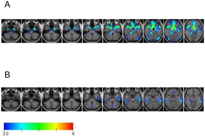
Data were collected prospectively on 28 elderly patients scheduled for elective cardiac surgery. MRI of the brains of all patients were assessed for prior cerebral infarctions, and carotid and intracranial arterial stenosis. Patients also completed six neuropsychological tests of memory, attention and executive function before and after surgery. POCD was defined as an individual decrease in more than two tests of at least 1 standard deviation from the group baseline mean for that test. The degree of gray matter loss in the MTL of each patient was calculated using voxel-based morphometry with three-dimensional, T1-weighted MRI. This represented the degree of gray matter change as a Z score.
Postoperative cognitive dysfunction was identified in 8 of the 28 patients (29%). Patients with POCD had significantly more white matter lesions on MRI, and greater loss of gray matter in the bilateral MTL (average Z score 2.0±0.9) than patients without POCD. An analysis by stepwise logistic regression identified gray matter loss in the MTL and cerebral infarctions on MRI as independent predictors of POCD.
These preliminary findings suggested that reduced gray matter in the bilateral MTL and white matter lesions existed in brains of elderly cardiac surgery patients who experienced POCD. Additional studies with larger sample sizes are needed to confirm these findings.
术后认知功能障碍(POCD)被认为是老年患者心脏手术后的一种并发症。脑部成像为老年患者的神经退行性变提供了证据;然而,脑结构异常及其与POCD的关系尚不确定。这项初步研究调查了术前MRI显示的双侧内侧颞叶(MTL)灰质丢失是否与POCD有关。
前瞻性收集了28例计划进行择期心脏手术的老年患者的数据。对所有患者的脑部MRI进行评估,以确定是否存在先前的脑梗死、颈动脉和颅内动脉狭窄。患者还在手术前后完成了六项关于记忆、注意力和执行功能的神经心理学测试。POCD被定义为在至少两项测试中个体得分比该测试组基线平均值下降超过1个标准差。使用基于体素的形态测量法结合三维T1加权MRI计算每位患者MTL的灰质丢失程度。这以Z分数表示灰质变化程度。
28例患者中有8例(29%)出现术后认知功能障碍。与未发生POCD的患者相比,发生POCD的患者MRI上的白质病变明显更多,双侧MTL的灰质丢失也更严重(平均Z分数2.0±0.9)。逐步逻辑回归分析确定MTL灰质丢失和MRI上的脑梗死是POCD的独立预测因素。
这些初步研究结果表明,经历POCD的老年心脏手术患者大脑中存在双侧MTL灰质减少和白质病变。需要更多样本量的进一步研究来证实这些发现。