Liu Shixuan, Cheng Wei, Fowle Grider Ronald, Shen Guomin, Li Weikai
1] Department of Biochemistry and Molecular Biophysics, Washington University School of Medicine, St Louis, Missouri 63110, USA [2].
Department of Biochemistry and Molecular Biophysics, Washington University School of Medicine, St Louis, Missouri 63110, USA.
Nat Commun. 2014;5:3110. doi: 10.1038/ncomms4110.
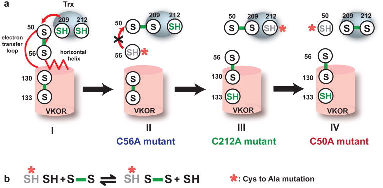
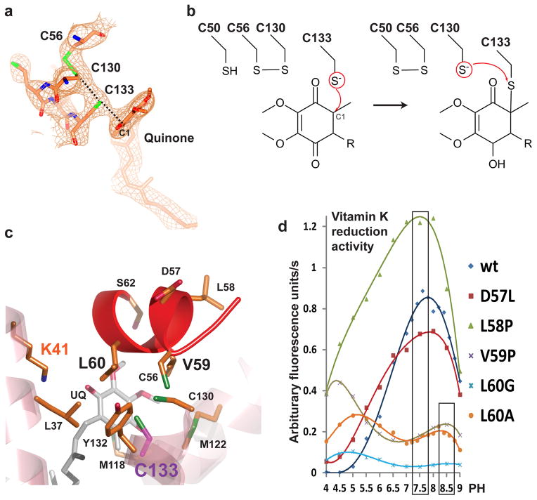
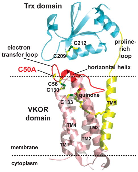
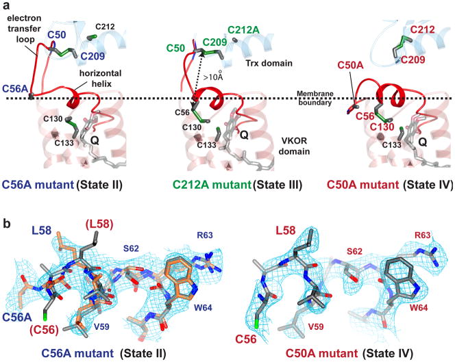
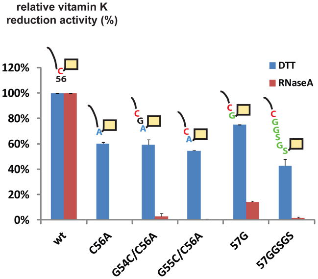
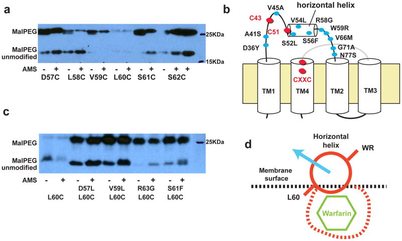
The intramembrane vitamin K epoxide reductase (VKOR) supports blood coagulation in humans and is the target of the anticoagulant warfarin. VKOR and its homologues generate disulphide bonds in organisms ranging from bacteria to humans. Here, to better understand the mechanism of VKOR catalysis, we report two crystal structures of a bacterial VKOR captured in different reaction states. These structures reveal a short helix at the hydrophobic active site of VKOR that alters between wound and unwound conformations. Motions of this 'horizontal helix' promote electron transfer by regulating the positions of two cysteines in an adjacent loop. Winding of the helix separates these 'loop cysteines' to prevent backward electron flow. Despite these motions, hydrophobicity at the active site is maintained to facilitate VKOR catalysis. Biochemical experiments suggest that several warfarin-resistant mutations act by changing the conformation of the horizontal helix. Taken together, these studies provide a comprehensive understanding of VKOR function.
膜内维生素K环氧化物还原酶(VKOR)维持人体血液凝固,是抗凝血剂华法林的作用靶点。VKOR及其同源物在从细菌到人类的各种生物体中生成二硫键。在此,为了更好地理解VKOR催化机制,我们报告了处于不同反应状态的细菌VKOR的两种晶体结构。这些结构揭示了VKOR疏水活性位点处的一个短螺旋,其在缠绕和未缠绕构象之间变化。这个“水平螺旋”的运动通过调节相邻环中两个半胱氨酸的位置来促进电子转移。螺旋的缠绕使这些“环半胱氨酸”分开,以防止电子逆向流动。尽管有这些运动,活性位点的疏水性得以维持以促进VKOR催化。生化实验表明,几种对华法林耐药的突变是通过改变水平螺旋的构象起作用的。综上所述,这些研究提供了对VKOR功能的全面理解。