Tytgat Institute for Liver and Intestinal Research, Academic Medical Center (AMC), Amsterdam, The Netherlands.
Department of Gastroenterology, University Hospital Leuven, Catholic University of Leuven, Leuven, Belgium.
PLoS One. 2014 Jan 29;9(1):e87785. doi: 10.1371/journal.pone.0087785. eCollection 2014.
Electrical stimulation of the vagus nerve suppresses intestinal inflammation and normalizes gut motility in a mouse model of postoperative ileus. The exact anatomical interaction between the vagus nerve and the intestinal immune system remains however a matter of debate. In the present study, we provide additional evidence on the direct and indirect vagal innervation of the spleen and analyzed the anatomical evidence for neuroimmune modulation of macrophages by vagal preganglionic and enteric postganglionic nerve fibers within the intestine.
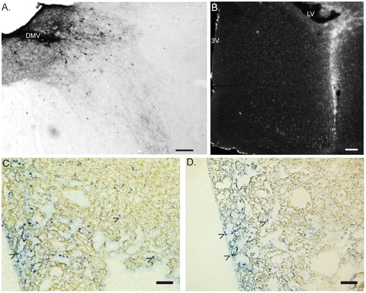
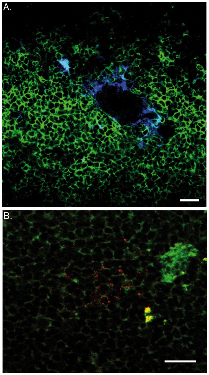
Dextran conjugates were used to label vagal preganglionic (motor) fibers projecting to the small intestine and spleen. Moreover, identification of the neurochemical phenotype of the vagal efferent fibers and enteric neurons was performed by immunofluorescent labeling. F4/80 antibody was used to label resident macrophages.
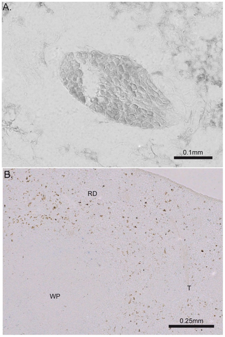
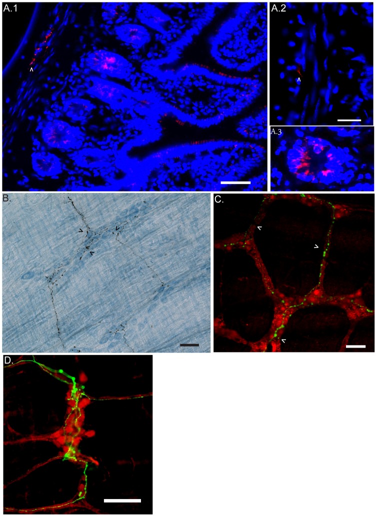
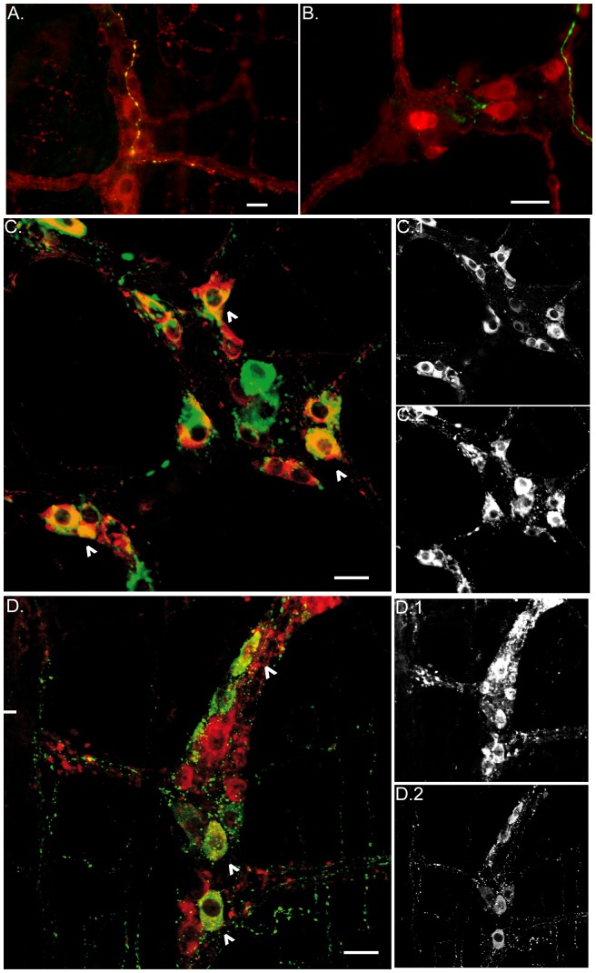
Our anterograde tracing experiments did not reveal dextran-labeled vagal fibers or terminals in the mesenteric ganglion or spleen. Vagal efferent fibers were confined within the myenteric plexus region of the small intestine and mainly endings around nNOS, VIP and ChAT positive enteric neurons. nNOS, VIP and ChAT positive fibers were found in close proximity of intestinal resident macrophages carrying α7 nicotinic receptors. Of note, VIP receptors were found on resident macrophages located in close proximity of VIP positive nerve fibers.
In the present study, we show that the vagus nerve does not directly interact with resident macrophages in the gut or spleen. Instead, the vagus nerve preferentially interacts with nNOS, VIP and ChAT enteric neurons located within the gut muscularis with nerve endings in close proximity of the resident macrophages.
电刺激迷走神经可抑制术后肠麻痹模型中小鼠的肠道炎症并使肠道运动正常化。然而,迷走神经与肠道免疫系统之间的确切解剖学相互作用仍存在争议。在本研究中,我们提供了迷走神经对脾的直接和间接支配的更多证据,并分析了迷走节前和肠节后神经纤维在肠道内对巨噬细胞的神经免疫调节的解剖学证据。
使用葡聚糖缀合物标记投射到小肠和脾脏的迷走节前(运动)纤维。此外,通过免疫荧光标记鉴定迷走传出纤维和肠神经元的神经化学表型。使用 F4/80 抗体标记驻留巨噬细胞。
我们的顺行示踪实验未显示迷走神经纤维或末端在肠系膜神经节或脾脏中被标记。迷走传出纤维局限于小肠的肌间神经丛区域,主要围绕 nNOS、VIP 和 ChAT 阳性肠神经元的末端。在携带α7 烟碱受体的肠道驻留巨噬细胞附近发现了 nNOS、VIP 和 ChAT 阳性纤维。值得注意的是,在 VIP 阳性神经纤维附近的驻留巨噬细胞上发现了 VIP 受体。
在本研究中,我们表明迷走神经不会直接与肠道或脾脏中的驻留巨噬细胞相互作用。相反,迷走神经优先与位于肠道肌层内的 nNOS、VIP 和 ChAT 肠神经元相互作用,其末端与驻留巨噬细胞接近。