Yoneyama Masanori, Shiba Tatsuo, Hasebe Shigeru, Umeda Kasumi, Yamaguchi Taro, Ogita Kiyokazu
Department of Pharmacology, Setsunan University Faculty of Pharmaceutical Sciences, Hirakata, Osaka, Japan.
PLoS One. 2014 Feb 4;9(2):e87953. doi: 10.1371/journal.pone.0087953. eCollection 2014.
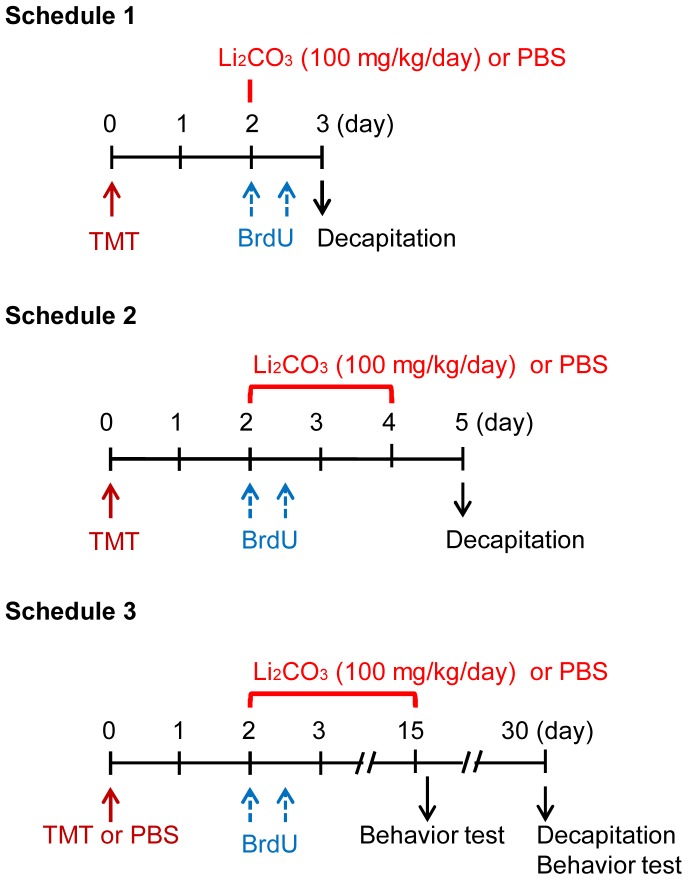
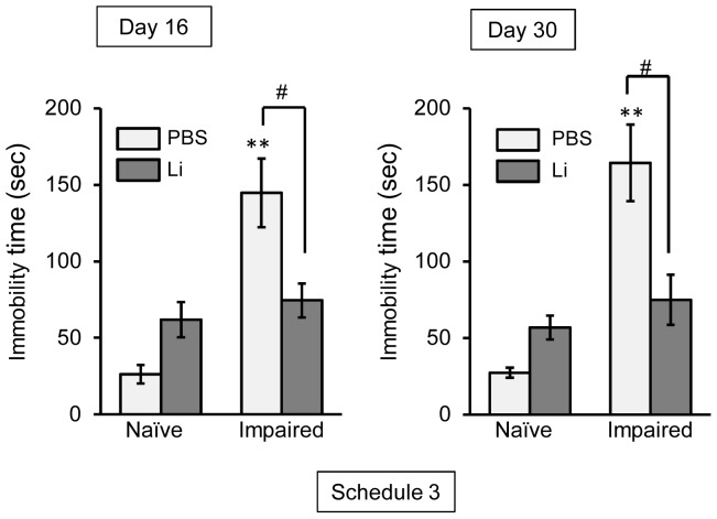
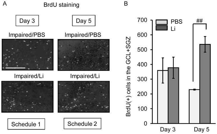
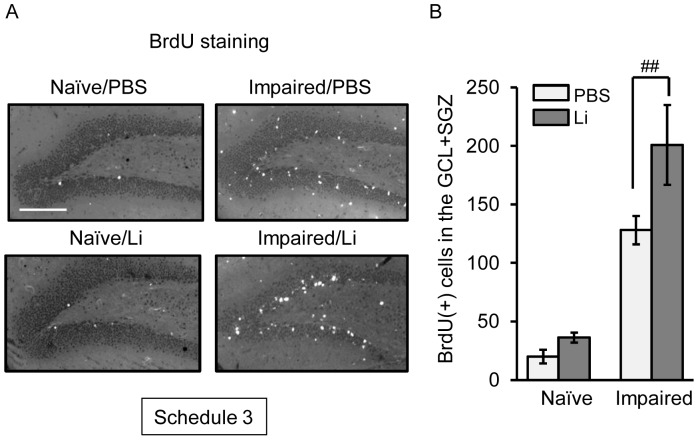
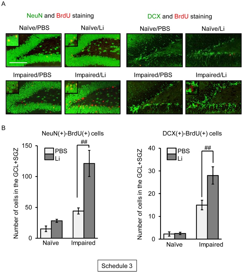
Lithium, a mood stabilizer, is known to ameliorate the stress-induced decrease in hippocampal neurogenesis seen in animal models of stress-related disorders. However, it is unclear whether lithium has beneficial effect on neuronal repair following neuronal damage in neuronal degenerative diseases. Here, we evaluated the effect of in vivo treatment with lithium on the hippocampal neuronal repair in a mouse model of trimethyltin (TMT)-induced neuronal loss/self-repair in the hippocampal dentate gyrus (such mice referred to as "impaired animals") [Ogita et al. (2005) J Neurosci Res 82: 609-621]. The impaired animals had a dramatically increased number of 5-bromo-2'-deoxyuridine (BrdU)-incorporating cells in their dentate gyrus at the initial time window (days 3 to 5 post-TMT treatment) of the self-repair stage. A single treatment with lithium produced no significant change in the number of BrdU-incorporating cells in the dentate granule cell layer and subgranular zone on day 3 post-TMT treatment. On day 5 post-TMT treatment, however, BrdU-incorporating cells were significantly increased in number by lithium treatment for 3 days. Most interestingly, chronic treatment (15 days) with lithium increased the number of BrdU-incorporating cells positive for NeuN or doublecortin in the dentate granule cell layer of the impaired animals, but not in that of naïve animals. The results of a forced swimming test revealed that the chronic treatment with lithium improved the depression-like behavior seen in the impaired animals. Taken together, our data suggest that lithium had a beneficial effect on neuronal repair following neuronal loss in the dentate gyrus through promoted proliferation and survival/neuronal differentiation of neural stem/progenitor cells in the subgranular zone.
锂作为一种情绪稳定剂,已知其可改善应激相关障碍动物模型中所见的应激诱导的海马神经发生减少。然而,尚不清楚锂对神经退行性疾病中神经元损伤后的神经元修复是否具有有益作用。在此,我们评估了在三甲基锡(TMT)诱导的海马齿状回神经元丢失/自我修复小鼠模型(此类小鼠称为“受损动物”)中体内给予锂对海马神经元修复的影响[小田等人(2005年)《神经科学研究杂志》82:609 - 621]。在自我修复阶段的初始时间窗口(TMT处理后3至5天),受损动物齿状回中5-溴-2'-脱氧尿苷(BrdU)掺入细胞的数量显著增加。在TMT处理后第3天,单次给予锂对齿状颗粒细胞层和颗粒下区中BrdU掺入细胞的数量没有产生显著变化。然而,在TMT处理后第5天,锂处理3天使BrdU掺入细胞的数量显著增加。最有趣的是,锂的慢性处理(15天)增加了受损动物齿状颗粒细胞层中NeuN或双皮质素阳性的BrdU掺入细胞的数量,但在未处理动物中未增加。强迫游泳试验的结果表明,锂的慢性处理改善了受损动物中出现的抑郁样行为。综上所述,我们的数据表明,锂通过促进颗粒下区神经干/祖细胞的增殖、存活/神经元分化,对齿状回神经元丢失后的神经元修复具有有益作用。