Shah Claudio, Hegde Balachandra G, Morén Björn, Behrmann Elmar, Mielke Thorsten, Moenke Gregor, Spahn Christian M T, Lundmark Richard, Daumke Oliver, Langen Ralf
Max-Delbrück-Center for Molecular Medicine, Crystallography, Robert-Rössle-Straße 10, 13092 Berlin, Germany.
Institute of Chemistry and Biochemistry, Free University Berlin, Takustraße 6, 14195 Berlin, Germany.
Structure. 2014 Mar 4;22(3):409-420. doi: 10.1016/j.str.2013.12.015. Epub 2014 Feb 6.
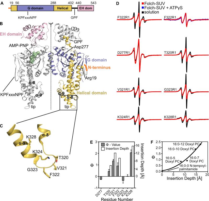
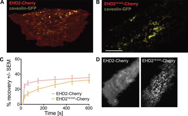
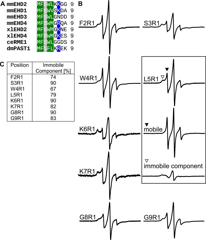
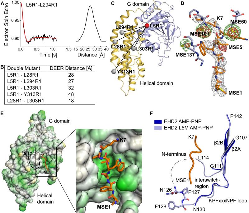
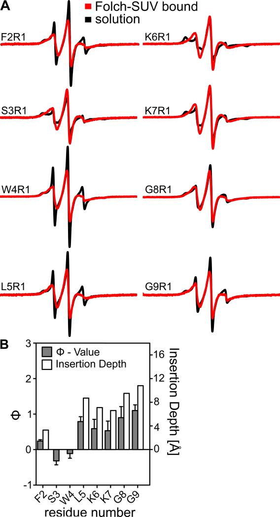
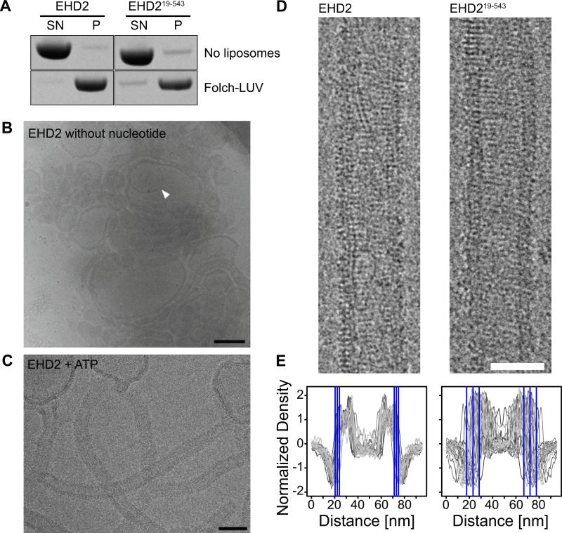
The dynamin-related Eps15-homology domain-containing protein 2 (EHD2) is a membrane-remodeling ATPase that regulates the dynamics of caveolae. Here, we established an electron paramagnetic resonance (EPR) approach to characterize structural features of membrane-bound EHD2. We show that residues at the tip of the helical domain can insert into the membrane and may create membrane curvature by a wedging mechanism. Using EPR and X-ray crystallography, we found that the N terminus is folded into a hydrophobic pocket of the GTPase domain in solution and can be released into the membrane. Cryoelectron microscopy demonstrated that the N terminus is not essential for oligomerization of EHD2 into a membrane-anchored scaffold. Instead, we found a function of the N terminus in regulating targeting and stable association of EHD2 to caveolae. Our data uncover an unexpected, membrane-induced regulatory switch in EHD2 and demonstrate the versatility of EPR to study structure and function of dynamin superfamily proteins.
动力蛋白相关的含Eps15同源结构域蛋白2(EHD2)是一种调节小窝动力学的膜重塑ATP酶。在此,我们建立了一种电子顺磁共振(EPR)方法来表征膜结合型EHD2的结构特征。我们发现,螺旋结构域末端的残基可插入膜中,并可能通过楔入机制产生膜曲率。利用EPR和X射线晶体学,我们发现N端在溶液中折叠成GTP酶结构域的疏水口袋,并可释放到膜中。冷冻电子显微镜显示,N端对于EHD2寡聚形成膜锚定支架并非必不可少。相反,我们发现N端在调节EHD2靶向小窝并与之稳定结合方面具有功能。我们的数据揭示了EHD2中一个意想不到的膜诱导调节开关,并证明了EPR在研究动力蛋白超家族蛋白结构和功能方面的多功能性。