Bach Martin, Schimmelpfennig Christoph, Stolzing Alexandra
Fraunhofer Institute for Cell Therapy and Immunology, Leipzig, Germany ; Universitätsklinik Leipzig, Department für Innere Medizin, Neurologie und Dermatologie, Sektion Rheumatologie, Leipzig, Germany.
Fraunhofer Institute for Cell Therapy and Immunology, Leipzig, Germany ; Klinikum St. Georg, Klinik für Internistische Onkologie und Hämatologie, Leipzig, Germany.
PLoS One. 2014 Feb 6;9(2):e88115. doi: 10.1371/journal.pone.0088115. eCollection 2014.
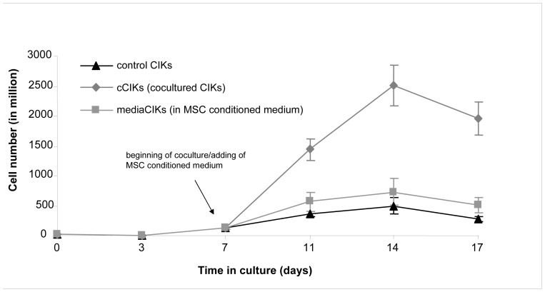
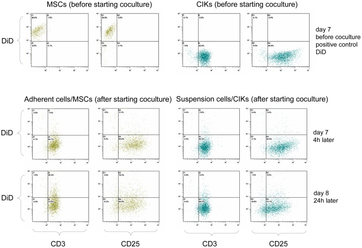
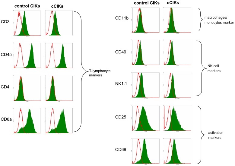
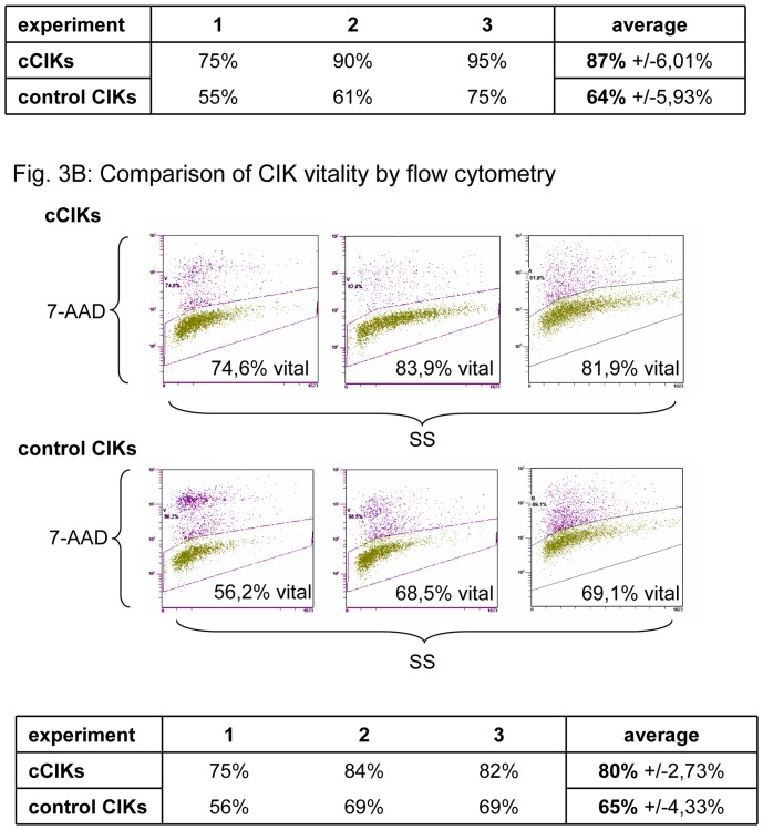
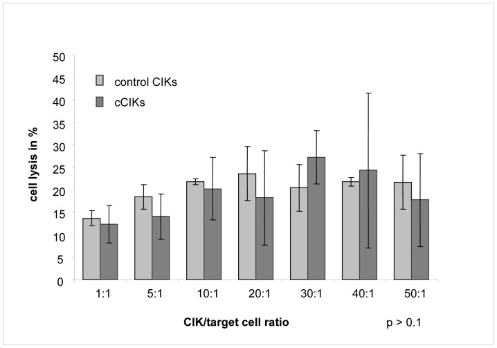
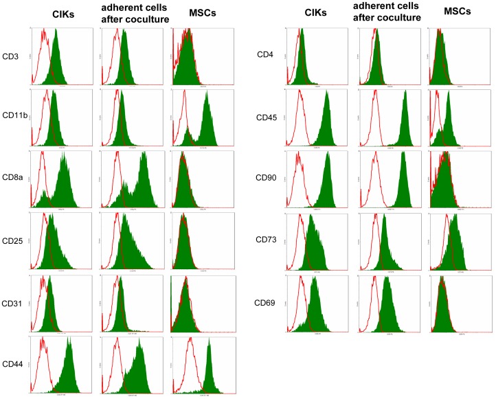
Stimulating lymphocytes with Ifn-γ, anti-CD3, and interleukin-2 promotes the proliferation of a cell population coexpressing T-lymphocyte surface antigens such as CD3, CD8a, and CD25 as well as natural killer cell markers such as NK1.1, CD49, and CD69. These cells, referred to as cytokine-induced killer cells (CIKs), display cytotoxic activity against tumour cells, even without prior antigen presentation, and offer a new cell-based approach to the treatment of malignant diseases. Because CIKs are limited in vivo, strategies to optimize in vitro culture yield are required. In the last 10 years, mesenchymal stem cells (MSCs) have gathered considerable attention. Aside from their uses in tissue engineering and as support in haematopoietic stem cell transplantations, MSCs show notable immunomodulatory characteristics, providing further possibilities for therapeutic applications. In this study, we investigated the influence of murine MSCs on proliferation, phenotype, vitality, and cytotoxicity of murine CIKs in a coculture system. We found that CIKs in coculture proliferated within 7 days, with an average growth factor of 18.84, whereas controls grew with an average factor of 3.7 in the same period. Furthermore, higher vitality was noted in cocultured CIKs than in controls. Cell phenotype was unaffected by coculture with MSCs and, notably, coculture did not impact cytotoxicity against the tumour cells analysed. The findings suggest that cell-cell contact is primarily responsible for these effects. Humoral interactions play only a minor role. Furthermore, no phenotypical MSCs were detected after coculture for 4 h, suggesting the occurrence of immune reactions between CIKs and MSCs. Further investigations with DiD-labelled MSCs revealed that the observed disappearance of MSCs appears not to be due to differentiation processes.
用干扰素-γ、抗CD3和白细胞介素-2刺激淋巴细胞可促进共表达T淋巴细胞表面抗原(如CD3、CD8a和CD25)以及自然杀伤细胞标志物(如NK1.1、CD49和CD69)的细胞群体增殖。这些细胞被称为细胞因子诱导的杀伤细胞(CIK),即使没有预先的抗原呈递,也对肿瘤细胞具有细胞毒性活性,并为恶性疾病的治疗提供了一种新的基于细胞的方法。由于CIK在体内数量有限,需要优化体外培养产量的策略。在过去10年中,间充质干细胞(MSC)受到了广泛关注。除了在组织工程中的应用以及作为造血干细胞移植的支持外,MSC还具有显著的免疫调节特性,为治疗应用提供了更多可能性。在本研究中,我们在共培养系统中研究了小鼠MSC对小鼠CIK增殖、表型、活力和细胞毒性的影响。我们发现,共培养中的CIK在7天内增殖,平均生长因子为18.84,而同期对照组的平均生长因子为3.7。此外,共培养的CIK的活力高于对照组。细胞表型不受与MSC共培养的影响,值得注意的是,共培养不影响对所分析肿瘤细胞的细胞毒性。研究结果表明,细胞间接触是这些效应的主要原因。体液相互作用仅起次要作用。此外,共培养4小时后未检测到表型MSC,这表明CIK与MSC之间发生了免疫反应。用DiD标记的MSC进行的进一步研究表明,观察到的MSC消失似乎不是由于分化过程。