• 文献检索
  • 文档翻译
  • 深度研究
  • 学术资讯
  • Suppr Zotero 插件Zotero 插件
  • 邀请有礼
  • 套餐&价格
  • 历史记录
应用&插件
Suppr Zotero 插件Zotero 插件浏览器插件Mac 客户端Windows 客户端微信小程序
定价
高级版会员购买积分包购买API积分包
服务
文献检索文档翻译深度研究API 文档MCP 服务
关于我们
关于 Suppr公司介绍联系我们用户协议隐私条款
关注我们

Suppr 超能文献

核心技术专利:CN118964589B侵权必究
粤ICP备2023148730 号-1Suppr @ 2026

文献检索

告别复杂PubMed语法,用中文像聊天一样搜索,搜遍4000万医学文献。AI智能推荐,让科研检索更轻松。

立即免费搜索

文件翻译

保留排版,准确专业,支持PDF/Word/PPT等文件格式,支持 12+语言互译。

免费翻译文档

深度研究

AI帮你快速写综述,25分钟生成高质量综述,智能提取关键信息,辅助科研写作。

立即免费体验

无色素预测:一种用于非色素性皮肤恶性肿瘤的决策算法。

Prediction without Pigment: a decision algorithm for non-pigmented skin malignancy.

机构信息

School of Medicine, The University of Queensland, Australia.

Department of Dermatology, Division of General Dermatology, Medical University of Vienna, Austria.

出版信息

Dermatol Pract Concept. 2014 Jan 31;4(1):59-66. doi: 10.5826/dpc.0401a09. eCollection 2014 Jan.

DOI:10.5826/dpc.0401a09
PMID:24520516
原文链接:https://pmc.ncbi.nlm.nih.gov/articles/PMC3919842/
Abstract

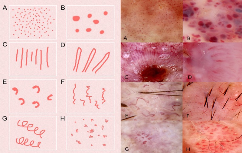
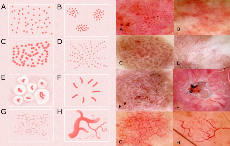
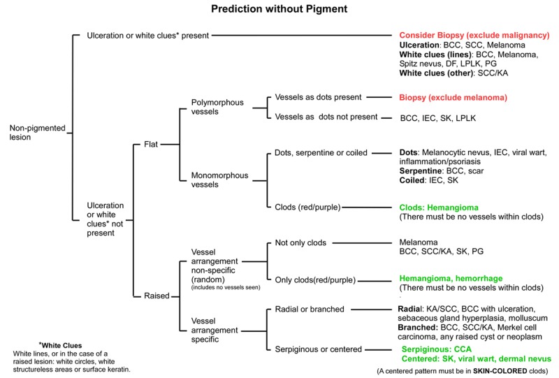
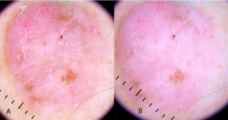
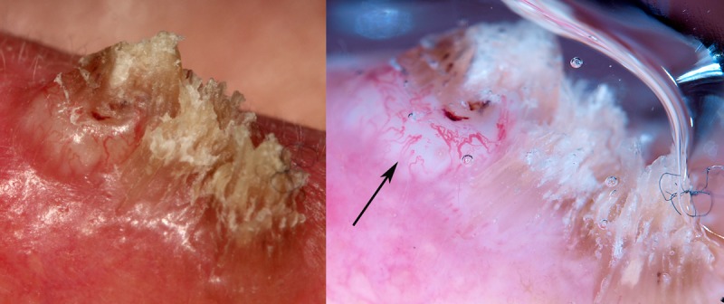
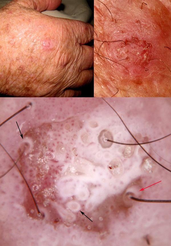
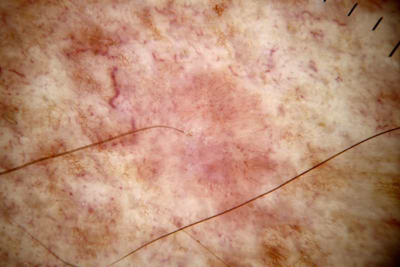
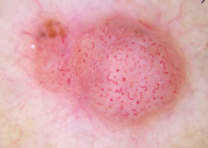
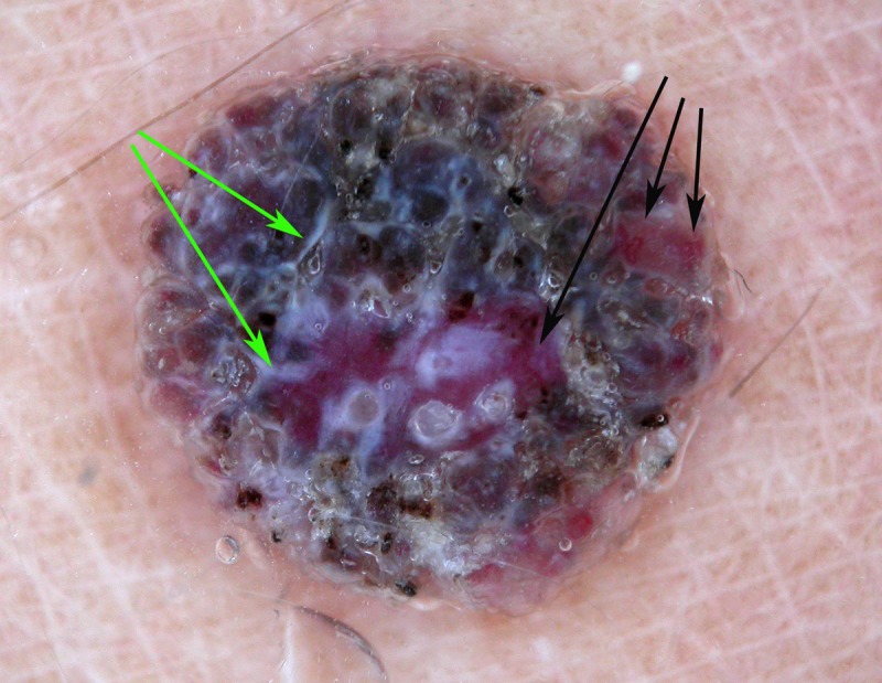
While there are several published comprehensive stepwise algorithmic methods for diagnosing pigmented skin malignancy, only limited material has been published for the stepwise assessment of non-pigmented lesions. We present a method based on pattern analysis, with a stepwise assessment, first, for ulceration, second, for white clues (defined as white lines, or in the case of a raised lesion any of the keratin clues: dermatoscopic white circles, dermatoscopic white structureless areas or surface keratin), and third, if no ulceration or white clues are present, proceed to vessel pattern analysis. This is a novel method, and apart from the assessment of white clues in raised lesions, it has not been formally tested. The priority of keratin clues in raised lesions over vessel pattern analysis has, however, been verified. It is conceded that this method is less specific than methods which have clues of pigmented structures, and accepting these limitations, Prediction without Pigment is a decision algorithm intended to guide the clinician in the decision as to whether to perform a biopsy rather than consistently leading to a specific diagnosis. Reaching a more specific diagnosis at the end of our flowchart can be achieved by weighing of clues both clinical and dermatoscopic, and that ability can be expected to improve with both knowledge and experience, but no diagnostic method, including this one, can be 100% sensitive in diagnosing malignancy, in particular, melanoma. Taking these limitations into account, any non-pigmented lesion, regardless of pattern analysis, which is raised and firm (nodular) and for which a confident, specific benign diagnosis cannot be made, should be excised to exclude the nodular variant of amelanotic melanoma.

摘要

虽然已经有几个发表的用于诊断色素性皮肤恶性肿瘤的全面逐步算法方法,但只有有限的材料被发表用于逐步评估非色素病变。我们提出了一种基于模式分析的方法,首先进行溃疡评估,其次是白色线索(定义为白线,或者在隆起病变的情况下,任何角化线索:皮肤镜下的白色圆圈、皮肤镜下无结构的白色区域或表面角化),如果没有溃疡或白色线索,则进行血管模式分析。这是一种新的方法,除了评估隆起病变中的白色线索外,它尚未经过正式测试。然而,角化线索在隆起病变中的优先级已经得到了验证。需要承认的是,与具有色素结构线索的方法相比,这种方法的特异性较低,在接受这些局限性的前提下,无色素预测是一种决策算法,旨在指导临床医生决定是否进行活检,而不是始终导致特定的诊断。通过对临床和皮肤镜下的线索进行权衡,可以在我们的流程图的最后达到更具体的诊断,并且随着知识和经验的积累,这种能力有望提高,但没有任何诊断方法,包括这种方法,能够 100%敏感地诊断恶性肿瘤,特别是黑色素瘤。考虑到这些局限性,任何非色素性病变,无论模式分析如何,如果是隆起且坚硬(结节状),并且无法做出明确的良性诊断,都应切除以排除无色素性黑素瘤的结节变体。

https://cdn.ncbi.nlm.nih.gov/pmc/blobs/9878/3919842/c4d705e89b81/dp0401a09g010.jpg
https://cdn.ncbi.nlm.nih.gov/pmc/blobs/9878/3919842/5d6e6464f7fe/dp0401a09g001.jpg
https://cdn.ncbi.nlm.nih.gov/pmc/blobs/9878/3919842/625337480c99/dp0401a09g002.jpg
https://cdn.ncbi.nlm.nih.gov/pmc/blobs/9878/3919842/aa228e144d0c/dp0401a09g003.jpg
https://cdn.ncbi.nlm.nih.gov/pmc/blobs/9878/3919842/8521ed1457dd/dp0401a09g004.jpg
https://cdn.ncbi.nlm.nih.gov/pmc/blobs/9878/3919842/0a44dd03fe35/dp0401a09g005.jpg
https://cdn.ncbi.nlm.nih.gov/pmc/blobs/9878/3919842/f6f64a7398a5/dp0401a09g006.jpg
https://cdn.ncbi.nlm.nih.gov/pmc/blobs/9878/3919842/df774ea40e1a/dp0401a09g007.jpg
https://cdn.ncbi.nlm.nih.gov/pmc/blobs/9878/3919842/2f1b9b79112e/dp0401a09g008.jpg
https://cdn.ncbi.nlm.nih.gov/pmc/blobs/9878/3919842/c8f0af6e23f9/dp0401a09g009.jpg
https://cdn.ncbi.nlm.nih.gov/pmc/blobs/9878/3919842/c4d705e89b81/dp0401a09g010.jpg

相似文献

1
Prediction without Pigment: a decision algorithm for non-pigmented skin malignancy.无色素预测:一种用于非色素性皮肤恶性肿瘤的决策算法。
Dermatol Pract Concept. 2014 Jan 31;4(1):59-66. doi: 10.5826/dpc.0401a09. eCollection 2014 Jan.
2
Role of In Vivo Reflectance Confocal Microscopy in the Analysis of Melanocytic Lesions.体内反射共聚焦显微镜在黑素细胞性病变分析中的作用
Acta Dermatovenerol Croat. 2018 Apr;26(1):64-67.
3
Nodular melanoma: five consecutive cases in a general practice with polarized and non-polarized dermatoscopy and dermatopathology.结节性黑素瘤:在一家普通诊所中,通过偏振和非偏振皮肤镜检术及皮肤病理学连续观察到的五例病例。
Dermatol Pract Concept. 2014 Apr 30;4(2):69-75. doi: 10.5826/dpc.0402a15. eCollection 2014 Apr.
4
Dermatoscopy of amelanotic and hypomelanotic melanoma.无色素性和色素减退性黑色素瘤的皮肤镜检查
J Dtsch Dermatol Ges. 2014 Jun;12(6):467-72. doi: 10.1111/ddg.12368. Epub 2014 May 14.
5
Dermatoscopy of flat pigmented facial lesions.面部扁平色素性皮损的皮肤镜检查
J Eur Acad Dermatol Venereol. 2015 Jan;29(1):120-7. doi: 10.1111/jdv.12483. Epub 2014 Mar 24.
6
Dermatoscopic finding of white circles in an invasive pigmented melanoma.侵袭性色素性黑素瘤中出现白色圆圈的皮肤镜表现。
Australas J Dermatol. 2023 May;64(2):e202-e203. doi: 10.1111/ajd.13985. Epub 2023 Jan 30.
7
Dermatoscopy of Neoplastic Skin Lesions: Recent Advances, Updates, and Revisions.皮肤肿瘤性病变的皮肤镜检查:最新进展、更新和修订。
Curr Treat Options Oncol. 2018 Sep 20;19(11):56. doi: 10.1007/s11864-018-0573-6.
8
Dermatoscopic findings and dermatopathological correlates in clinical variants of actinic keratosis, Bowen's disease, keratoacanthoma, and squamous cell carcinoma.光化性角化病、鲍恩病、角化棘皮瘤和鳞状细胞癌的临床变异型的皮肤镜表现和皮肤病理相关性。
Dermatol Ther. 2021 May;34(3):e14877. doi: 10.1111/dth.14877. Epub 2021 Mar 7.
9
The reticular point of view in dermatoscopy.皮肤镜检查中的网状观点。
J Am Acad Dermatol. 2009 Oct;61(4):605-10. doi: 10.1016/j.jaad.2009.04.006. Epub 2009 Aug 22.
10
Dermatoscopic features of pigmented and non-pigmented basal cell carcinoma.色素性和非色素性基底细胞癌的皮肤镜特征。
Georgian Med News. 2015 Jan(238):50-3.

引用本文的文献

1
Unseen Aggressor: A Case of Amelanotic Melanoma With Orbital Metastasis and Imaging-Pathology Correlation.隐匿性侵袭者:一例伴有眼眶转移的无色素性黑色素瘤病例及影像-病理相关性分析
Cureus. 2025 Jul 19;17(7):e88333. doi: 10.7759/cureus.88333. eCollection 2025 Jul.
2
Dermoscopic Features of Eccrine Poromas in Diverse Skin Phototypes: A Retrospective Study of 26 Cases.不同皮肤光型中汗腺汗孔瘤的皮肤镜特征:一项26例病例的回顾性研究
Dermatol Pract Concept. 2025 Apr 1;15(2):5071. doi: 10.5826/dpc.1502a5071.
3
Cutaneous Metastasis of Triple Negative Breast Cancer: A Case Report and Review of Literature.

本文引用的文献

1
Dermoscopy of squamous cell carcinoma and keratoacanthoma.鳞状细胞癌和角化棘皮瘤的皮肤镜检查
Arch Dermatol. 2012 Dec;148(12):1386-92. doi: 10.1001/archdermatol.2012.2974.
2
Dermoscopic criteria and basal cell carcinoma.皮肤镜检查标准与基底细胞癌。
G Ital Dermatol Venereol. 2012 Apr;147(2):135-40.
3
White shiny structures in melanoma and BCC.黑色素瘤和基底细胞癌中的白色发亮结构。
三阴性乳腺癌的皮肤转移:一例报告及文献复习
Cureus. 2025 Mar 2;17(3):e79926. doi: 10.7759/cureus.79926. eCollection 2025 Mar.
4
Trauma-Associated Pseudo-Hutchinson Sign: An Autobiographical Case Report Emphasizing Conditions, Pseudo-Conditions, and Pseudo-Pseudo-Conditions.创伤相关假性哈钦森征:一份强调情况、假性情况和假假性情况的病例报告
Cureus. 2024 Dec 27;16(12):e76474. doi: 10.7759/cureus.76474. eCollection 2024 Dec.
5
Amelanotic Melanoma-Biochemical and Molecular Induction Pathways.无色素性黑素瘤的生化和分子诱导途径。
Int J Mol Sci. 2024 Oct 26;25(21):11502. doi: 10.3390/ijms252111502.
6
Thin Amelanotic and Hypomelanotic Melanoma: Clinicopathological and Dermoscopic Features.薄型非色素性和低色素性黑素瘤:临床病理和皮肤镜特征。
Medicina (Kaunas). 2024 Jul 30;60(8):1239. doi: 10.3390/medicina60081239.
7
Patients poorly recognize lesions of concern that are malignant melanomas: is self-screening the correct advice?患者无法识别恶性黑色素瘤等有问题的病变:自我筛查是正确的建议吗?
PeerJ. 2024 Jul 3;12:e17674. doi: 10.7717/peerj.17674. eCollection 2024.
8
Implementation of artificial intelligence for the detection of cutaneous melanoma within a primary care setting: prevalence and types of skin cancer in outdoor enthusiasts.在初级保健环境中实施人工智能检测皮肤黑色素瘤:户外爱好者的皮肤癌患病率和类型。
PeerJ. 2023 Aug 8;11:e15737. doi: 10.7717/peerj.15737. eCollection 2023.
9
Point prevalence of non-melanoma and melanoma skin cancers in Australian surfers and swimmers in Southeast Queensland and Northern New South Wales.昆士兰州东南部和新南威尔士州北部澳大利亚冲浪者和游泳者中非黑色素瘤和黑色素瘤皮肤癌的现患率。
PeerJ. 2022 Apr 28;10:e13243. doi: 10.7717/peerj.13243. eCollection 2022.
10
Cutaneous metastases of embryonal rhabdomyosarcoma in a black African child: the role of dermoscopy in reducing time to and cost of diagnosis.一名非洲黑人儿童胚胎性横纹肌肉瘤的皮肤转移:皮肤镜检查在缩短诊断时间和降低诊断成本方面的作用
Ecancermedicalscience. 2021 Oct 12;15:1304. doi: 10.3332/ecancer.2021.1304. eCollection 2021.
Arch Dermatol. 2012 Jan;148(1):146. doi: 10.1001/archdermatol.2011.618.
4
The significance of crystalline/chrysalis structures in the diagnosis of melanocytic and nonmelanocytic lesions.结晶/蛹期结构在黑素细胞和非黑素细胞病变诊断中的意义。
J Am Acad Dermatol. 2012 Aug;67(2):194.e1-8. doi: 10.1016/j.jaad.2011.04.039. Epub 2011 Oct 24.
5
Dermatoscopy of facial actinic keratosis, intraepidermal carcinoma, and invasive squamous cell carcinoma: a progression model.面部光化性角化病、表皮内癌和侵袭性鳞状细胞癌的皮肤镜检查:一个进展模型。
J Am Acad Dermatol. 2012 Apr;66(4):589-97. doi: 10.1016/j.jaad.2011.02.011. Epub 2011 Aug 11.
6
Diagnostic accuracy of dermatoscopy for melanocytic and nonmelanocytic pigmented lesions.皮肤镜检查对黑素细胞和非黑素细胞色素性皮损的诊断准确性。
J Am Acad Dermatol. 2011 Jun;64(6):1068-73. doi: 10.1016/j.jaad.2010.03.039. Epub 2011 Mar 25.
7
How to diagnose nonpigmented skin tumors: a review of vascular structures seen with dermoscopy: part II. Nonmelanocytic skin tumors.如何诊断非色素性皮肤肿瘤:皮肤镜下所见血管结构的综述:第二部分。非黑素细胞性皮肤肿瘤。
J Am Acad Dermatol. 2010 Sep;63(3):377-86; quiz 387-8. doi: 10.1016/j.jaad.2009.11.697.
8
Dermatoscopy of basal cell carcinoma: morphologic variability of global and local features and accuracy of diagnosis.基底细胞癌的皮肤镜检查:整体和局部特征的形态变异性及诊断准确性。
J Am Acad Dermatol. 2010 Jan;62(1):67-75. doi: 10.1016/j.jaad.2009.05.035. Epub 2009 Oct 13.
9
New dermoscopic pattern in actinic keratosis and related conditions.光化性角化病及相关病症中的新皮肤镜模式。
Arch Dermatol. 2009 Jun;145(6):732. doi: 10.1001/archdermatol.2009.86.
10
Observation of chrysalis structures with polarized dermoscopy.偏振皮肤镜下蝶蛹结构的观察
Arch Dermatol. 2009 May;145(5):618. doi: 10.1001/archdermatol.2009.28.