Zhu Xiaohua, Li Jinbo, Hong Yeongjin, Kimura Richard H, Ma Xiaowei, Liu Hongguang, Qin Chunxia, Hu Xiang, Hayes Thomas R, Benny Paul, Gambhir Sanjiv Sam, Cheng Zhen
Molecular Imaging Program at Stanford (MIPS), Department of Radiology, and Bio-X Program, Canary Center at Stanford for Cancer Early Detection, Stanford University , Stanford, California 94305-5344, United States.
Mol Pharm. 2014 Apr 7;11(4):1208-17. doi: 10.1021/mp400683q. Epub 2014 Feb 24.
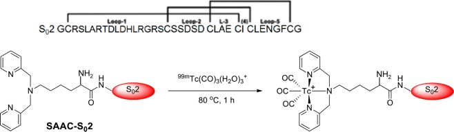
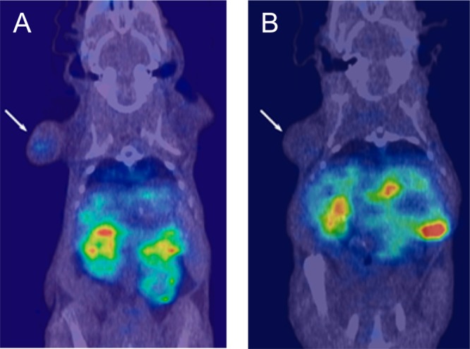
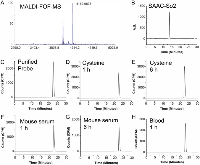
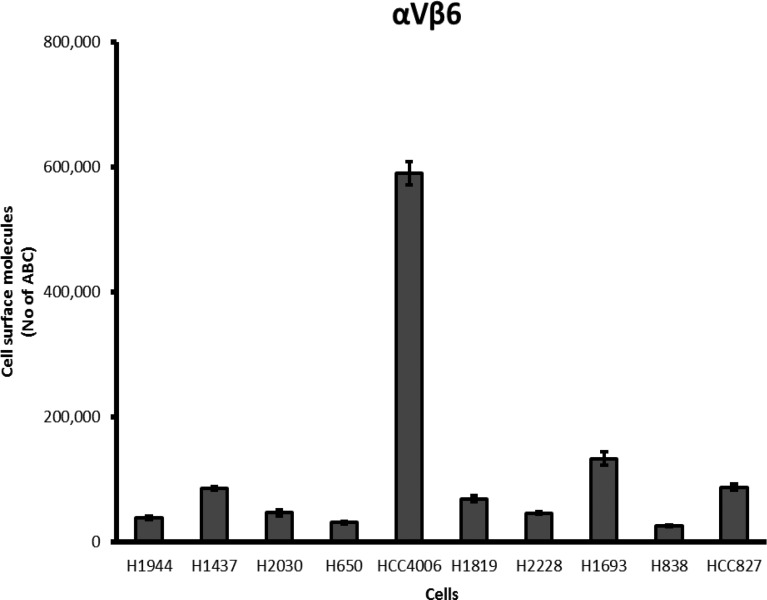
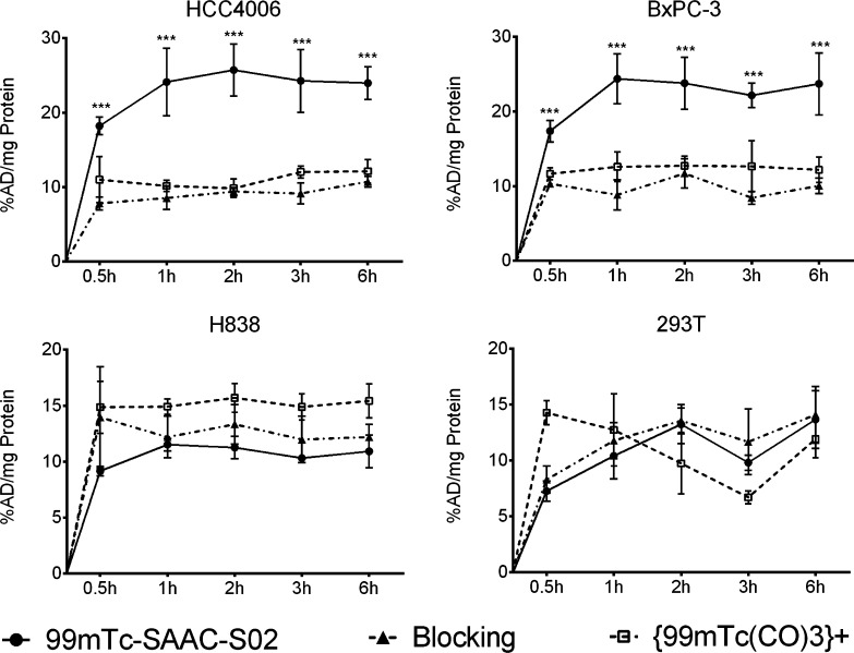
Integrin αvβ6 is overexpressed in a variety of cancers, and its expression is often associated with poor prognosis. Therefore, there is a need to develop affinity reagents for noninvasive imaging of integrin αvβ6 expression since it may provide early cancer diagnosis, more accurate prognosis, and better treatment planning. We recently engineered and validated highly stable cystine knot peptides that selectively bind integrin αvβ6 with no cross-reactivity to integrins αvβ5, α5β1, or αvβ3, also known to be overexpressed in many cancers. Here, we developed a single photon emission computed tomography (SPECT) probe for imaging integrin αvβ6 positive tumors. Cystine knot peptide, S02, was first conjugated with a single amino acid chelate (SAAC) and labeled with (99m)Tc(H2O)3(CO)3. The resulting probe, (99m)Tc-SAAC-S02, was then evaluated by in vitro cell uptake studies using two αvβ6 positive cell lines (human lung adenocarcinoma cell line HCC4006 and pancreatic cancer cell line BxPC-3) and two αvβ6 negative cell lines (human lung adenocarcinoma cell line H838 and human embryonic kidney cell line 293T). Next, SPECT/CT and biodistribution studies were performed in nude mice bearing HCC4006 and H838 tumor xenografts to evaluate the in vivo performance of (99m)Tc-SAAC-S02. Significant differences in the uptake of (99m)Tc-SAAC-S02 were observed in αvβ6 positive vs negative cells (P < 0.05). Biodistribution and small animal SPECT/CT studies revealed that (99m)Tc-SAAC-S02 accumulated to moderate levels in antigen positive tumors (∼2% ID/g at 1 and 6 h postinjection, n = 3 or 4/group). Moreover, the probe demonstrated tumor-to-background tissue ratios of 6.81 ± 2.32 (tumor-to-muscle) and 1.63 ± 0.18 (tumor-to-blood) at 6 h postinjection in αvβ6 positive tumor xenografts. Co-incubation of the probe with excess amount of unlabeled S02 as a blocking agent demonstrated significantly reduced tumor uptake, which is consistent with specific binding to the target. Renal filtration was the main route of clearance. In conclusion, knottin peptides are excellent scaffolds for which to develop highly stable imaging probes for a variety of oncological targets. (99m)Tc-SAAC-S02 demonstrates promise for use as a SPECT agent to image integrin αvβ6 expression in living systems.
整合素αvβ6在多种癌症中过度表达,其表达通常与不良预后相关。因此,需要开发用于整合素αvβ6表达无创成像的亲和试剂,因为它可能提供早期癌症诊断、更准确的预后和更好的治疗方案。我们最近设计并验证了高度稳定的胱氨酸结肽,其能选择性结合整合素αvβ6,对整合素αvβ5、α5β1或αvβ3无交叉反应,这些整合素在许多癌症中也已知会过度表达。在此,我们开发了一种用于成像整合素αvβ6阳性肿瘤的单光子发射计算机断层扫描(SPECT)探针。首先将胱氨酸结肽S02与单氨基酸螯合剂(SAAC)偶联,并用(99m)Tc(H2O)3(CO)3标记。然后使用两种αvβ6阳性细胞系(人肺腺癌细胞系HCC4006和胰腺癌细胞系BxPC-3)和两种αvβ6阴性细胞系(人肺腺癌细胞系H838和人胚肾细胞系293T),通过体外细胞摄取研究对所得探针(99m)Tc-SAAC-S02进行评估。接下来,在携带HCC-4006和H838肿瘤异种移植的裸鼠中进行SPECT/CT和生物分布研究,以评估(99m)Tc-SAAC-S02的体内性能。在αvβ6阳性与阴性细胞中观察到(99m)Tc-SAAC-S02摄取存在显著差异(P<0.05)。生物分布和小动物SPECT/CT研究表明,(99m)Tc-SAAC-S02在抗原阳性肿瘤中积累到中等水平(注射后1小时和6小时约为2% ID/g,每组n = 3或4)。此外,在αvβ6阳性肿瘤异种移植中,注射后6小时该探针的肿瘤与背景组织比率为6.81±2.32(肿瘤与肌肉)和1.63±0.18(肿瘤与血液)。将探针与过量未标记的S02作为阻断剂共同孵育,显示肿瘤摄取显著降低,这与与靶标的特异性结合一致。肾脏滤过是主要的清除途径。总之,结蛋白肽是开发针对多种肿瘤靶点的高度稳定成像探针的优秀支架。(99m)Tc-SAAC-S02有望用作SPECT试剂,用于在活体系统中成像整合素αvβ6的表达。