Han Bo, Chen Szu-Yu, Zhu Ying-Ting, Tseng Scheffer C G
Department of Ophthalmology, Union Hospital, Tongji Medical College, Huazhong University of Science and Technology, Wuhan, PR China; Ocular Surface Research & Education Foundation, Miami, FL, USA.
R&D Department, TissueTech, Inc., Miami, FL, USA.
Stem Cell Res. 2014 Mar;12(2):562-73. doi: 10.1016/j.scr.2014.01.003. Epub 2014 Jan 22.
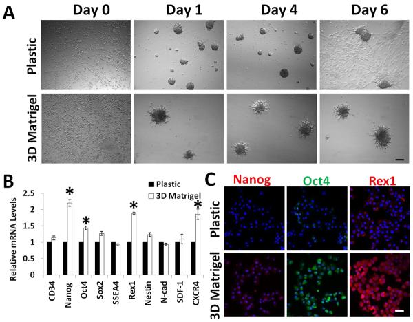
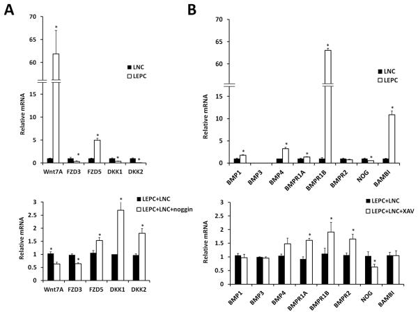
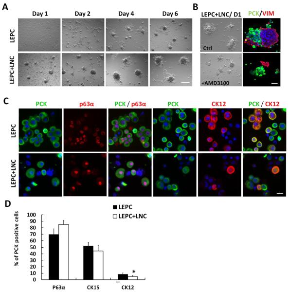
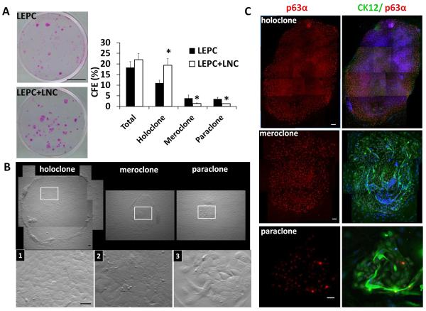
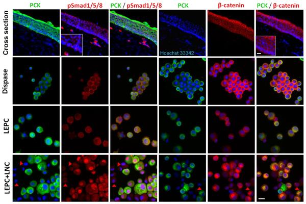
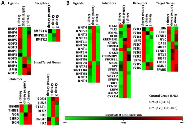
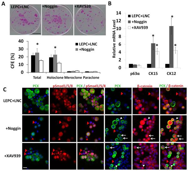
Both BMP and Wnt signaling control stem cells in bulge/dermal papilla, intestinal crypt, and bone marrow. To explore their roles in the limbal niche, which govern corneal epithelial homeostasis, we established an in vitro model of sphere growth by reunion between single limbal epithelial progenitor cells (LEPCs) and aggregates of limbal niche cells (LNCs) in 3D Matrigel. Compared to LEPCs alone, spheres formed by LEPC+LNC exhibited higher clonal growth and less corneal epithelial differentiation. Furthermore, pSmad1/5/8 was in the nucleus of LEPCs, but not LNCs, and correlated with upregulation of BMP1, BMP3, BMP4, all three BMP receptors, and BMP target genes. Inactivation of BMP signaling in LNCs was correlated with upregulation of noggin preferentially expressed by LNCs. Additionally, β-catenin was stabilized in the perinuclear cytoplasm in LEPCs and correlated with upregulation of Wnt7A and FZD5 preferentially expressed by LEPCs. Inactivation of Wnt signaling in LNCs was correlated with upregulation of DKK1/2 by LNCs. Addition of XAV939 that expectedly downregulated perinuclear β-catenin in LEPCs led to significant reduction of epithelial clonal growth, but upregulated all three BMP receptors and downregulated LNC-derived noggin, resulting in activation of BMP signaling in LNCs. Addition of noggin that expectedly downregulated nuclear localization of pSmad1/5/8 in LEPCs led to nuclear localization of β-catenin in larger LEPCs but membrane relocation of β-catenin in smaller LEPCs and significant upregulation of DKK1/2. Hence, balancing acts between Wnt signaling and BMP signaling exist not only within LEPCs but also between LEPCs and LNCs to regulate clonal growth of LEPCs.
骨形态发生蛋白(BMP)信号和Wnt信号均调控毛囊隆突部/毛乳头、肠隐窝和骨髓中的干细胞。为了探究它们在维持角膜上皮稳态的角膜缘生态位中的作用,我们通过在三维基质胶中将单个角膜缘上皮祖细胞(LEPC)与角膜缘生态位细胞(LNC)聚集体重聚,建立了一个球体生长的体外模型。与单独的LEPC相比,LEPC + LNC形成的球体表现出更高的克隆生长能力和更低的角膜上皮分化程度。此外,磷酸化的Smad1/5/8位于LEPC的细胞核中,而不在LNC的细胞核中,并且与BMP1、BMP3、BMP4、所有三种BMP受体以及BMP靶基因的上调相关。LNC中BMP信号的失活与LNC优先表达的头蛋白(noggin)的上调相关。此外,β-连环蛋白在LEPC的核周细胞质中稳定存在,并且与LEPC优先表达的Wnt7A和FZD5的上调相关。LNC中Wnt信号的失活与LNC中DKK1/2的上调相关。添加预期会下调LEPC核周β-连环蛋白的XAV939导致上皮克隆生长显著减少,但上调了所有三种BMP受体并下调了LNC来源的头蛋白,从而激活了LNC中的BMP信号。添加预期会下调LEPC中磷酸化Smad1/5/8核定位的头蛋白导致较大LEPC中β-连环蛋白的核定位,但较小LEPC中β-连环蛋白的膜转位以及DKK1/2的显著上调。因此,Wnt信号和BMP信号之间的平衡作用不仅存在于LEPC内部,也存在于LEPC和LNC之间,以调节LEPC的克隆生长。