Institute of Laser Engineering, Beijing University of Technology, Beijing 100124, China.
Nanoscale Res Lett. 2014 Feb 19;9(1):87. doi: 10.1186/1556-276X-9-87.
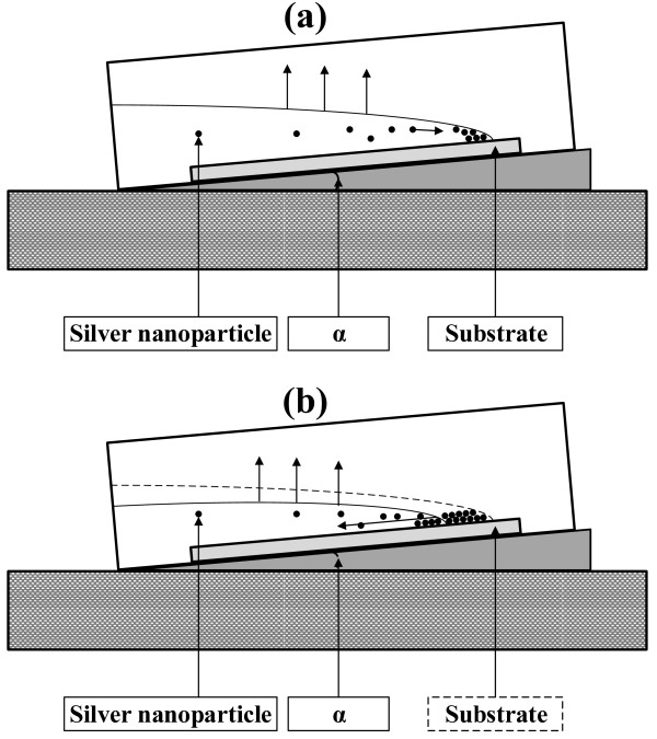
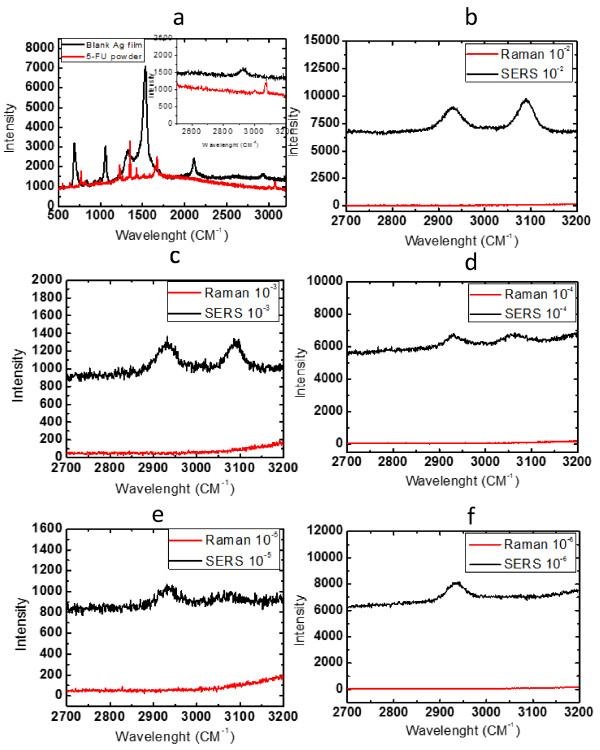
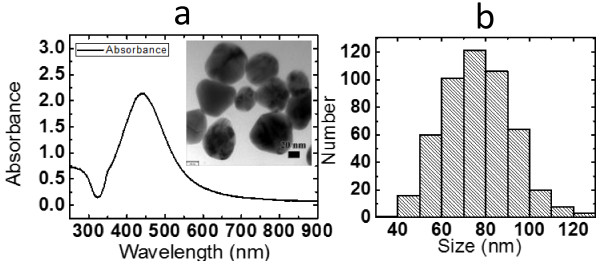
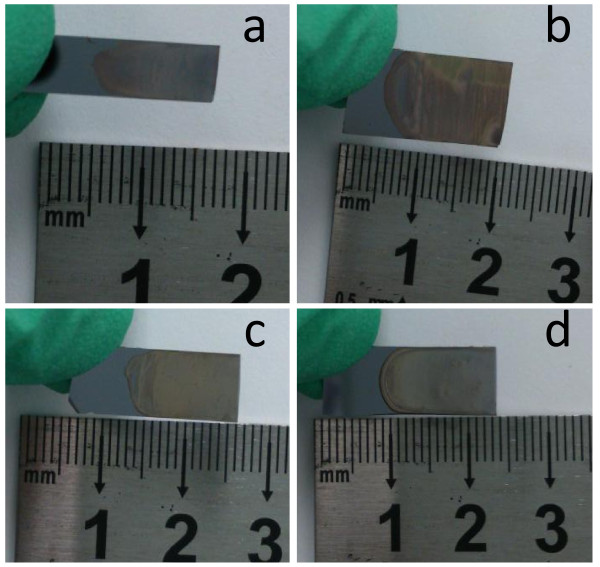
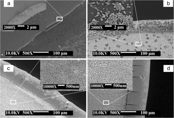
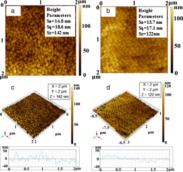
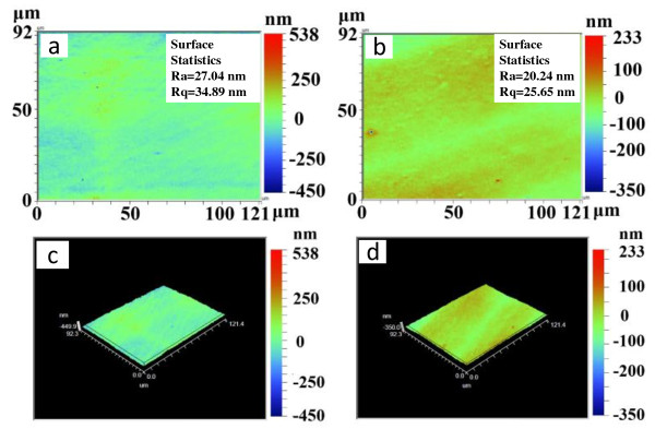
We report here a simple and innovative method to prepare large-scale silver nanoparticle films based on the controlled coffee ring effect. It is demonstrated that the films can be used as surface-enhanced Raman scattering probes to detect low-concentration medicines. Silver nanoparticles with the average size about 70 nm were prepared by reduction of silver nitride. In our experiment, the coffee ring effect was controlled by tilting the substrates during the deposition of silver nanoparticle films. Silver nanoparticle films were spontaneously formed on the surface of silicon substrates at the temperatures about 50°C based on the solvent evaporation and the coffee ring effect. The microstructure of the films was investigated using the scanning electron microscope and atomic force microscope. The surface roughness of the films is found as small as 20 nm. Then, the films were exposed to aqueous solutions of medicine at different concentrations. A comparison with a Raman spectra measured with a conventional Raman spectrometer showed that the Raman signal can be detected in the solution with concentrations as low as 1 × 10⁻⁵ M, and the enhancement factor achieved by the silver nanoparticle film can at least reach to 1.08 × 10⁴. Our experimental results indicate that this technique is promising in the production of large-scale silver nanoparticle films for the surface-enhanced Raman scattering. These may be utilized in biochemical and trace analytical applications.
我们在这里报告了一种基于控制咖啡环效应制备大规模银纳米粒子薄膜的简单创新方法。结果表明,这些薄膜可用作表面增强拉曼散射探针来检测低浓度药物。通过还原氮化银制备了平均粒径约为 70nm 的银纳米粒子。在我们的实验中,通过在沉积银纳米粒子薄膜时倾斜基底来控制咖啡环效应。基于溶剂蒸发和咖啡环效应,银纳米粒子在 50°C 左右的温度下自发地在硅基底表面形成薄膜。使用扫描电子显微镜和原子力显微镜研究了薄膜的微观结构。发现薄膜的表面粗糙度低至 20nm。然后,将薄膜暴露于不同浓度的药物水溶液中。与常规拉曼光谱仪测量的拉曼光谱进行比较表明,在浓度低至 1×10⁻⁵M 的溶液中可以检测到拉曼信号,银纳米粒子薄膜的增强因子至少可以达到 1.08×10⁴。我们的实验结果表明,该技术在制备用于表面增强拉曼散射的大规模银纳米粒子薄膜方面具有广阔的应用前景。这些薄膜可用于生化和痕量分析应用。