Chilek Jennifer L, Wang Ruhung, Draper Rockford K, Pantano Paul
Department of Chemistry, ‡Department of Molecular and Cell Biology, and §The Alan G. MacDiarmid NanoTech Institute, The University of Texas at Dallas , Richardson, Texas 75080, United States.
Anal Chem. 2014 Mar 18;86(6):2882-7. doi: 10.1021/ac403827m. Epub 2014 Mar 7.
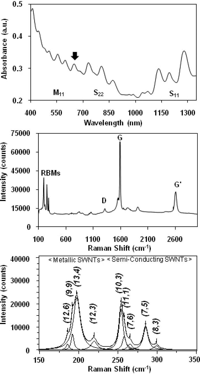
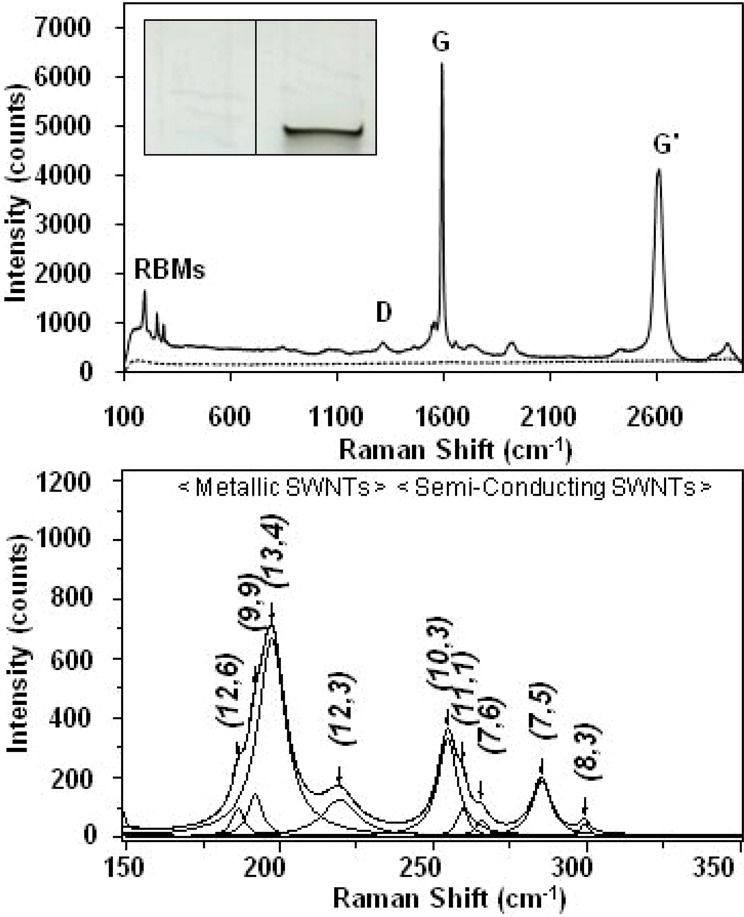
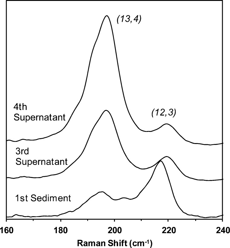
It is well-known that the uptake of single-walled carbon nanotubes (SWNTs) by living cells depends on factors such as SWNT length and surface chemistry. Surprisingly, little is known about whether the electronic structure of a SWNT influences uptake. One reason for this has been the lack of methods to measure the uptake of SWNTs by cell populations. Previously, we developed a rapid, sensitive, and label-free sodium dodecyl sulfate (SDS)-polyacrylamide gel electrophoresis (PAGE) method for measuring the amount of SWNTs in lysates prepared from cultured cells ( Wang et al. Anal. Chem. 2009 , 81 , 2944 ). Herein, we describe the use of SDS-PAGE and microprobe Raman spectroscopy to detect and distinguish the electronic structure of SWNTs internalized by mammalian cells. Using normal rat kidney (NRK) cells and SWNTs dispersed with bovine serum albumin (BSA), we demonstrate that the method can detect both metallic and semiconducting SWNTs in lysates of cells that had internalized BSA-SWNTs and that the uptake of BSA-SWNTs by NRK cells is not influenced by SWNT electronic structure.
众所周知,活细胞对单壁碳纳米管(SWNTs)的摄取取决于诸如SWNT长度和表面化学等因素。令人惊讶的是,关于SWNT的电子结构是否会影响摄取却知之甚少。造成这种情况的一个原因是缺乏测量细胞群体对SWNTs摄取的方法。此前,我们开发了一种快速、灵敏且无需标记的十二烷基硫酸钠(SDS)-聚丙烯酰胺凝胶电泳(PAGE)方法,用于测量从培养细胞制备的裂解物中SWNTs的含量(Wang等人,《分析化学》,2009年,81卷,2944页)。在此,我们描述了使用SDS-PAGE和微探针拉曼光谱来检测和区分哺乳动物细胞内化的SWNTs的电子结构。使用正常大鼠肾(NRK)细胞和用牛血清白蛋白(BSA)分散的SWNTs,我们证明该方法可以检测内化了BSA-SWNTs的细胞裂解物中的金属性和半导体性SWNTs,并且NRK细胞对BSA-SWNTs的摄取不受SWNT电子结构的影响。