Institute of Orthopedics, Shaanxi University of Traditional Chinese Medicine, Century Ave,, Xi'an 712000, PR China.
BMC Complement Altern Med. 2014 Feb 24;14:74. doi: 10.1186/1472-6882-14-74.
It has been suggested that the formation of osteoblasts in bone marrow is closely associated with adipogenesis, and the balance between osteogenesis and adipogenesis differentiation of MSCs (mesenchymal stem cells) is disrupted in osteoporosis. In order to improve the treatment of osteoporosis, available agents with roles of regulating the balance is highly desirable. Emodin is a natural anthraquinone derivative extracted from Chinese herbs, which have been used to treat bone diseases for thousands of years. However, the underlying molecular mechanisms of emodin in modulating osteogenesis and adipogenesis remain poorly understood.
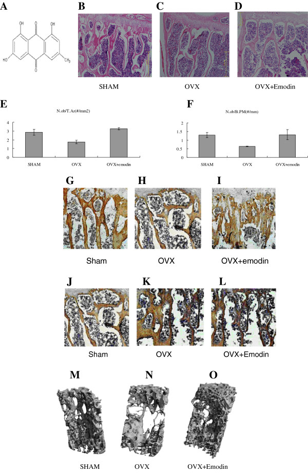
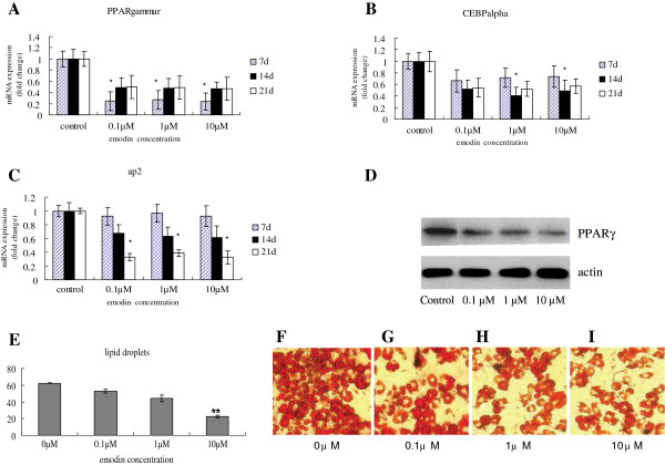
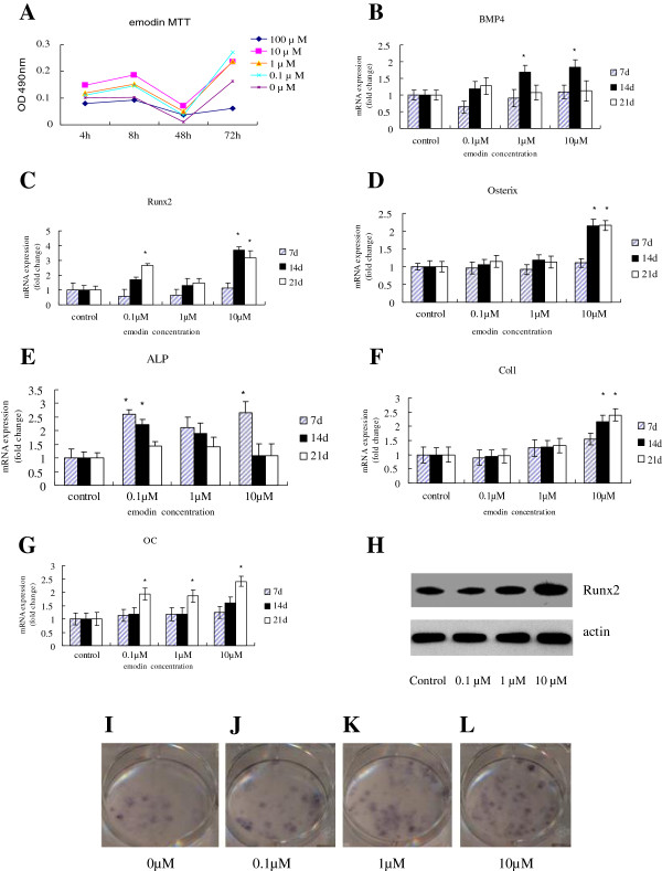
The molecular mechanisms of emodin on the processes of osteogenesis and adipogenesis in ovariectomized mouse and BMSCs (bone marrow mesenchymal stem cells) have been studied. We have analyzed the effects of emodin in vivo and in vitro. Female ICR mice were assigned to three groups: sham group, ovariectomy group, emodin group. Efficacy was evaluated by H&E, immunohistochemical assay and Micro-CT. In vitro, we analyze the effect of emodin--at concentrations between 0.1 μM and 10 μM--on the processes of inducing osteogenesis and inhibiting adipogenesis in BMSCs by ALP, Oil red O staining, real time RT-PCR and western blot.
As our experiment shows that emodin could increase the number of osteoblast, BMD (bone mineral density), BV/TV (trabecular bone volume fraction), Tb.N (trabecular number) and Conn.D (connectivity density) of OVX (ovariectomized) mice and decrease the bone marrow fat tissue and adipocytes. The genes and proteins expression of osteogenesis markers, such as Runx2, osterix, collagen type I, osteocalcin, or ALP were up-regulated. While, the genes and proteins involved in adipogenesis, PPARγ, C/EBPα and ap2 were down-regulated.
It proves that emodin inhibits adipocyte differentiation and enhances osteoblast differentiation from BMSCs.
据认为,骨髓中成骨细胞的形成与脂肪生成密切相关,而骨质疏松症中 MSC(间充质干细胞)的成骨和脂肪生成分化平衡被打破。为了改善骨质疏松症的治疗,具有调节平衡作用的现有药物是非常需要的。大黄素是一种从中药中提取的天然蒽醌衍生物,几千年来一直被用于治疗骨病。然而,大黄素调节成骨和脂肪生成的潜在分子机制仍知之甚少。
研究了大黄素在去卵巢小鼠和 BMSCs(骨髓间充质干细胞)成骨和脂肪生成过程中的分子机制。我们分析了大黄素在体内和体外的作用。将雌性 ICR 小鼠分为三组:假手术组、去卵巢组、大黄素组。通过 H&E、免疫组织化学检测和 Micro-CT 评估疗效。在体外,我们分析了大黄素(浓度在 0.1 μM 至 10 μM 之间)对 BMSCs 诱导成骨和抑制脂肪生成过程的影响,通过碱性磷酸酶(ALP)、油红 O 染色、实时 RT-PCR 和 Western blot 进行分析。
实验表明,大黄素可以增加去卵巢小鼠的成骨细胞数量、BMD(骨密度)、BV/TV(骨小梁体积分数)、Tb.N(骨小梁数量)和 Conn.D(连通密度),减少骨髓脂肪组织和脂肪细胞。成骨标志物基因和蛋白表达如 Runx2、osterix、I 型胶原、骨钙素或 ALP 上调。而参与脂肪生成的基因和蛋白,如 PPARγ、C/EBPα 和 ap2 下调。
证明大黄素抑制脂肪细胞分化,增强 BMSCs 成骨细胞分化。