Kumar Rajesh, Farahvar Salar, Ogren Jennifer A, Macey Paul M, Thompson Paul M, Woo Mary A, Yan-Go Frisca L, Harper Ronald M
Department of Anesthesiology, David Geffen School of Medicine at UCLA, University of California at Los Angeles, Los Angeles, CA 90095, USA ; Department of Radiological Sciences, David Geffen School of Medicine at UCLA, University of California at Los Angeles, Los Angeles, CA 90095, USA ; The Brain Research Institute, University of California at Los Angeles, Los Angeles, CA 90095, USA.
Department of Neurobiology, David Geffen School of Medicine at UCLA, University of California at Los Angeles, Los Angeles, CA 90095, USA.
Neuroimage Clin. 2014 Jan 31;4:383-91. doi: 10.1016/j.nicl.2014.01.009. eCollection 2014.
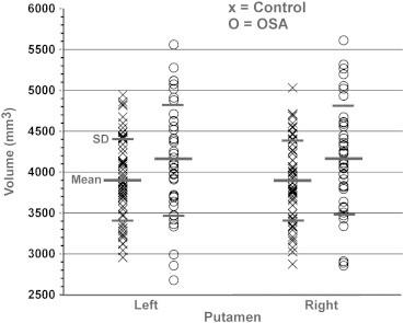
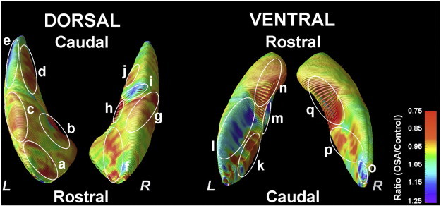
Obstructive sleep apnea (OSA) is accompanied by cognitive, motor, autonomic, learning, and affective abnormalities. The putamen serves several of these functions, especially motor and autonomic behaviors, but whether global and specific sub-regions of that structure are damaged is unclear. We assessed global and regional putamen volumes in 43 recently-diagnosed, treatment-naïve OSA (age, 46.4 ± 8.8 years; 31 male) and 61 control subjects (47.6 ± 8.8 years; 39 male) using high-resolution T1-weighted images collected with a 3.0-Tesla MRI scanner. Global putamen volumes were calculated, and group differences evaluated with independent samples t-tests, as well as with analysis of covariance (covariates; age, gender, and total intracranial volume). Regional differences between groups were visualized with 3D surface morphometry-based group ratio maps. OSA subjects showed significantly higher global putamen volumes, relative to controls. Regional analyses showed putamen areas with increased and decreased tissue volumes in OSA relative to control subjects, including increases in caudal, mid-dorsal, mid-ventral portions, and ventral regions, while areas with decreased volumes appeared in rostral, mid-dorsal, medial-caudal, and mid-ventral sites. Global putamen volumes were significantly higher in the OSA subjects, but local sites showed both higher and lower volumes. The appearance of localized volume alterations points to differential hypoxic or perfusion action on glia and other tissues within the structure, and may reflect a stage in progression of injury in these newly-diagnosed patients toward the overall volume loss found in patients with chronic OSA. The regional changes may underlie some of the specific deficits in motor, autonomic, and neuropsychologic functions in OSA.
阻塞性睡眠呼吸暂停(OSA)伴有认知、运动、自主神经、学习和情感异常。壳核参与其中多种功能,尤其是运动和自主神经行为,但该结构的整体及特定亚区域是否受损尚不清楚。我们使用3.0特斯拉MRI扫描仪采集的高分辨率T1加权图像,评估了43例新诊断的、未接受过治疗的OSA患者(年龄46.4±8.8岁;31例男性)和61例对照者(47.6±8.8岁;39例男性)的壳核整体及区域体积。计算壳核整体体积,并通过独立样本t检验以及协方差分析(协变量:年龄、性别和颅内总体积)评估组间差异。通过基于3D表面形态测量的组比率图可视化组间区域差异。与对照组相比,OSA患者的壳核整体体积显著更高。区域分析显示,与对照者相比,OSA患者壳核组织体积增加和减少的区域,包括尾侧、中背侧、中腹侧部分和腹侧区域体积增加,而体积减少的区域出现在嘴侧、中背侧、内侧尾侧和中腹侧部位。OSA患者的壳核整体体积显著更高,但局部部位体积有高有低。局部体积改变的出现表明该结构内的胶质细胞和其他组织受到不同的缺氧或灌注作用,可能反映了这些新诊断患者的损伤进展阶段,朝着慢性OSA患者中发现的总体积减少发展。区域变化可能是OSA患者运动、自主神经和神经心理功能某些特定缺陷的基础。