Kido Teruhito, Watanabe Kouki, Saeki Hideyuki, Shigemi Susumu, Matsuda Takeshi, Yamamoto Masaya, Kurata Akira, Kanza Rene Epunza, Itoh Toshihide, Mochizuki Teruhito
Department of Radiology, Ehime University, Toon, Japan.
Department of Cardiology, Saiseikai Matsuyama Hospital, Matsuyama, Japan.
Springerplus. 2014 Feb 7;3:75. doi: 10.1186/2193-1801-3-75. eCollection 2014.
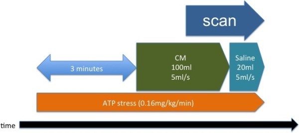
The purpose of this study was to investigate the utility incremental diagnostic value of combined assessment with coronary CT angiography (CCTA) and myocardial CT perfusion imaging (CTP) using dual-energy technology with an Adenosine Triphosphate (ATP) load technique.
Twenty-one patients underwent ATP-provocation dual-energy CT and CAG. We compared the diagnostic accuracy with CAG, for ischemic region due coronary stenosis by CCTA alone and CCTA combined with CTP (Combined CCTA/CTP).
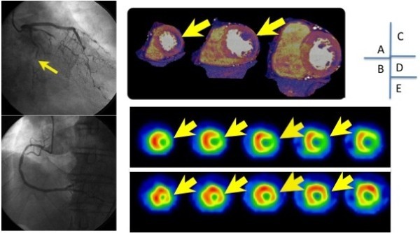
All of 21 patients CTP images could be evaluated, however 8 CCTA images could not be evaluated by calcification and motion artifact, so assessability was 61.9% (13/21) for CCTA alone, and 100% for Combined CCTA/CTP. With CAG results as a comparison, the sensitivity, specificity, positive predictive value, and negative predictive value were, respectively, 83.3% (20/24), 74.4% (29/39), 66.7% (20/30), and 87.8% (29/33) for CCTA alone, and 66.7% (16/24), 92.3% (36/39), 84.2% (16/19), and 81.8% (36/44) for combined CCTA/CTP. The diagnostic accuracy of the two methods were 77.8% (49/63) and 82.5% (52/63).
Dual-energy CT may be a useful modality for perfusion assessment and correlated well with the severity of stenosis on CAG. This technique may even be of use in cases of severe calcification in the coronary artery wall.
本研究旨在探讨采用三磷酸腺苷(ATP)负荷技术的双能技术联合冠状动脉CT血管造影(CCTA)和心肌CT灌注成像(CTP)的效用增量诊断价值。
21例患者接受了ATP激发双能CT和冠状动脉造影(CAG)。我们将单独使用CCTA以及CCTA联合CTP(联合CCTA/CTP)对因冠状动脉狭窄导致的缺血区域的诊断准确性与CAG进行了比较。
21例患者的CTP图像均可评估,但8例CCTA图像因钙化和运动伪影无法评估,因此单独CCTA的可评估性为61.9%(13/21),联合CCTA/CTP为100%。以CAG结果作为对照,单独CCTA的敏感性、特异性、阳性预测值和阴性预测值分别为83.3%(20/24)、74.4%(29/39)、66.7%(20/30)和87.8%(29/33),联合CCTA/CTP分别为66.7%(16/24)、92.3%(36/39)、84.2%(16/19)和81.8%(36/44)。两种方法的诊断准确性分别为77.8%(49/6)和82.5%(52/63)。
双能CT可能是一种用于灌注评估的有用方式,并且与CAG上的狭窄严重程度相关性良好。该技术甚至可能适用于冠状动脉壁严重钙化的病例。