Bajek Anna, Pokrywka Lukasz, Wolski Zbigniew, Dębski Robert, Drewa Tomasz
Department of Tissue Engineering, Nicolaus Copernicus University, Bydgoszcz, Poland.
Department of General, Oncological and Pediatric Urology, Nicolaus Copernicus University, Bydgoszcz, Poland.
Cent European J Urol. 2011;64(4):256-7. doi: 10.5173/ceju.2011.04.art15. Epub 2011 Dec 9.
Induction of apoptosis in prostatic epithelial cells by doxazosin, terazosin and prazosin has been well documented. However, the biochemical pathways of doxazosin action is still unclear. Aforementioned drugs should lead to decrease of prostate volume, although this effect was never observed in patients suffering from BPH after treatment with α1-antagonists. Probably, it is connected with cancer stem cells' resistance on chemotherapeutic agents. The aim of this study was to compare incidence of apoptosis induced by doxazosin in progenitor and differentiated cells isolated from human prostate epithelium.
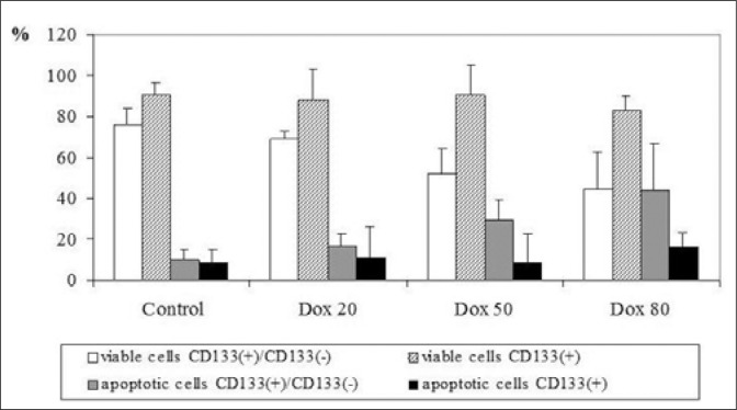
For this purpose tissue specimens were obtained from 10 patients suffering from BPH, the primary cultures of prostate epithelium were established and CD133 MicroBeads sorting was prepared. Both, CD133(+)/CD133(-) co-cultures and CD133(+) cells were incubated with different concentration of doxazosin for 12 h. Cell viability and apoptosis was estimated with Annexin V-FITC.
12 h incubation of CD133(+)/CD133(-) co-cultures with doxazosin resulted in increase of apoptotic cells, while in CD133(+) cultures no changes were observed. Correlation between apoptotic cell number and doxazosin concentration in CD133(+)/ CD133(-) co-cultures group was high (R = 0.99).
Doxazosin induced apoptosis in co-cultures of progenitor and differentiated epithelial cells. However, progenitor cells were not susceptible to apoptosis, what can be a reason of treatment failure in BPH patients.
多沙唑嗪、特拉唑嗪和哌唑嗪诱导前列腺上皮细胞凋亡已得到充分证实。然而,多沙唑嗪的作用生化途径仍不清楚。上述药物应会导致前列腺体积减小,尽管在用α1拮抗剂治疗良性前列腺增生(BPH)患者后从未观察到这种效果。这可能与癌症干细胞对化疗药物的抗性有关。本研究的目的是比较多沙唑嗪诱导人前列腺上皮中分离出的祖细胞和分化细胞凋亡的发生率。
为此,从10例BPH患者获取组织标本,建立前列腺上皮原代培养物,并制备CD133微珠分选。将CD133(+)/CD133(-)共培养物和CD133(+)细胞均与不同浓度的多沙唑嗪孵育12小时。用膜联蛋白V-异硫氰酸荧光素评估细胞活力和凋亡情况。
多沙唑嗪与CD133(+)/CD133(-)共培养物孵育12小时导致凋亡细胞增加,而在CD133(+)培养物中未观察到变化。CD133(+)/CD133(-)共培养物组中凋亡细胞数与多沙唑嗪浓度之间的相关性很高(R = 0.99)。
多沙唑嗪在祖细胞和分化上皮细胞的共培养物中诱导凋亡。然而,祖细胞不易发生凋亡,这可能是BPH患者治疗失败的原因。