Günther Jeannine, Kill Angela, Becker Mike Oliver, Heidecke Harald, Rademacher Judith, Siegert Elise, Radić Mislav, Burmester Gerd-Rüdiger, Dragun Duska, Riemekasten Gabriela
Arthritis Res Ther. 2014 Mar 11;16(2):R65. doi: 10.1186/ar4503.
Agonistic autoantibodies (Aabs) against the angiotensin II receptor type 1 (AT1R) and the endothelin receptor type A (ETAR) have been identified in patients with systemic sclerosis (SSc). In our present study, we examined the expression of the AT1R and the ETAR in human immune cells and the pathological effects mediated through these receptors by their corresponding Aabs.
Protein expression of AT1R and ETAR on peripheral blood mononuclear cells (PBMCs) from healthy individuals and SSc patients was analyzed using flow cytometry, and mRNA expression of both receptors in PBMCs from healthy donors was examined by real-time PCR. In addition, PBMCs from healthy donors were stimulated in vitro with affinity-purified immunoglobulin G (IgG) fractions from SSc patients positive for AT1R and ETAR Aabs, as well as with IgG from healthy donors serving as controls. Alterations in cell surface marker expression, cytokine secretion and chemotactic motility were analyzed using flow cytometry, enzyme-linked immunosorbent assays and chemotaxis assays, respectively. The results were correlated with the characteristics and clinical findings of the IgG donors.
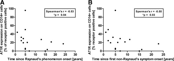
Both AT1R and ETAR were expressed on PBMCs in humans. Protein expression of both receptors was decreased in SSc patients compared with that of healthy donors and declined during the course of disease. IgG fractions of SSc patients positive for AT1R and ETAR Aabs induced T-cell migration in an Aab level-dependent manner. Moreover, IgG of SSc patients stimulated PBMCs to produce more interleukin 8 (IL-8) and chemokine (C-C motif) ligand 18 (CCL18) than did the IgG of healthy donors. All effects were significantly reduced by selective AT1R and ETAR antagonists. Statistical analysis revealed an association of SSc-IgG induced high IL-8 concentrations with an early disease stage and of high CCL18 concentrations with lung fibrosis onset and vascular complications in the respective IgG donors.
In our present study, we could demonstrate the expression of both AT1R and ETAR on human peripheral T cells, B cells and monocytes. The decreased receptor expression in SSc patients, the inflammatory and profibrotic effects upon Aab stimulation of PBMCs in vitro and the associations with clinical findings suggest a role for Aab-induced activation of immune cells mediated by the AT1R and the ETAR in the pathogenesis or even the onset of the disease.
在系统性硬化症(SSc)患者中已鉴定出针对1型血管紧张素II受体(AT1R)和A型内皮素受体(ETAR)的激动性自身抗体(Aab)。在本研究中,我们检测了AT1R和ETAR在人免疫细胞中的表达,以及相应Aab通过这些受体介导的病理效应。
使用流式细胞术分析健康个体和SSc患者外周血单个核细胞(PBMC)上AT1R和ETAR的蛋白表达,并通过实时PCR检测健康供体PBMC中这两种受体的mRNA表达。此外,用来自AT1R和ETAR Aab阳性的SSc患者的亲和纯化免疫球蛋白G(IgG)组分以及来自健康供体的IgG作为对照,体外刺激健康供体的PBMC。分别使用流式细胞术、酶联免疫吸附测定和趋化性测定分析细胞表面标志物表达、细胞因子分泌和趋化运动的变化。结果与IgG供体的特征和临床发现相关。
AT1R和ETAR均在人PBMC上表达。与健康供体相比,SSc患者中这两种受体的蛋白表达降低,且在疾病过程中下降。AT1R和ETAR Aab阳性的SSc患者的IgG组分以Aab水平依赖性方式诱导T细胞迁移。此外,SSc患者的IgG刺激PBMC产生的白细胞介素8(IL-8)和趋化因子(C-C基序)配体18(CCL18)比健康供体的IgG更多。选择性AT1R和ETAR拮抗剂可显著降低所有效应。统计分析显示,SSc-IgG诱导的高IL-8浓度与疾病早期相关,高CCL18浓度与相应IgG供体的肺纤维化发作和血管并发症相关。
在本研究中,我们能够证明AT1R和ETAR在人外周T细胞、B细胞和单核细胞上均有表达。SSc患者中受体表达降低、体外Aab刺激PBMC后的炎症和促纤维化作用以及与临床发现的相关性表明,Aab诱导的由AT1R和ETAR介导的免疫细胞激活在疾病的发病机制甚至疾病发作中起作用。