Suttkus A, Rohn S, Weigel S, Glöckner P, Arendt T, Morawski M
University of Leipzig, Department for Molecular and Cellular Mechanisms of Neurodegeneration, Paul Flechsig Institute for Brain Research, Jahnallee 59, Leipzig 04109, Germany.
1] University of Leipzig, Department for Molecular and Cellular Mechanisms of Neurodegeneration, Paul Flechsig Institute for Brain Research, Jahnallee 59, Leipzig 04109, Germany [2] SUNY Upstate Medical University, Department of Neuroscience and Physiology, WH 3240 750 East Adams Street, Syracuse, NY 13210, USA.
Cell Death Dis. 2014 Mar 13;5(3):e1119. doi: 10.1038/cddis.2014.25.
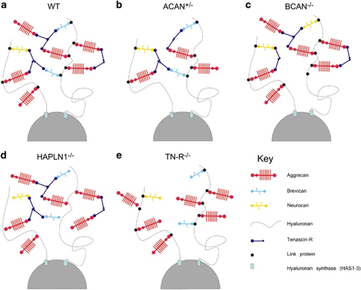
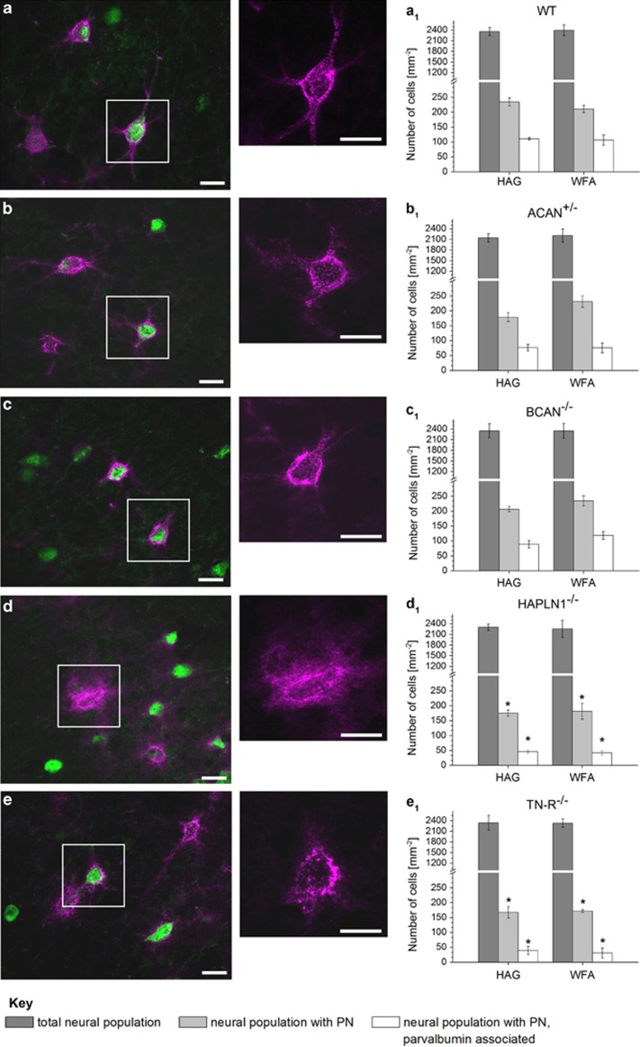
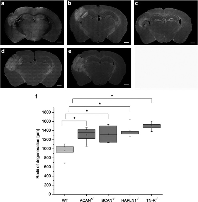
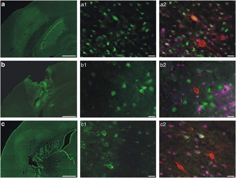
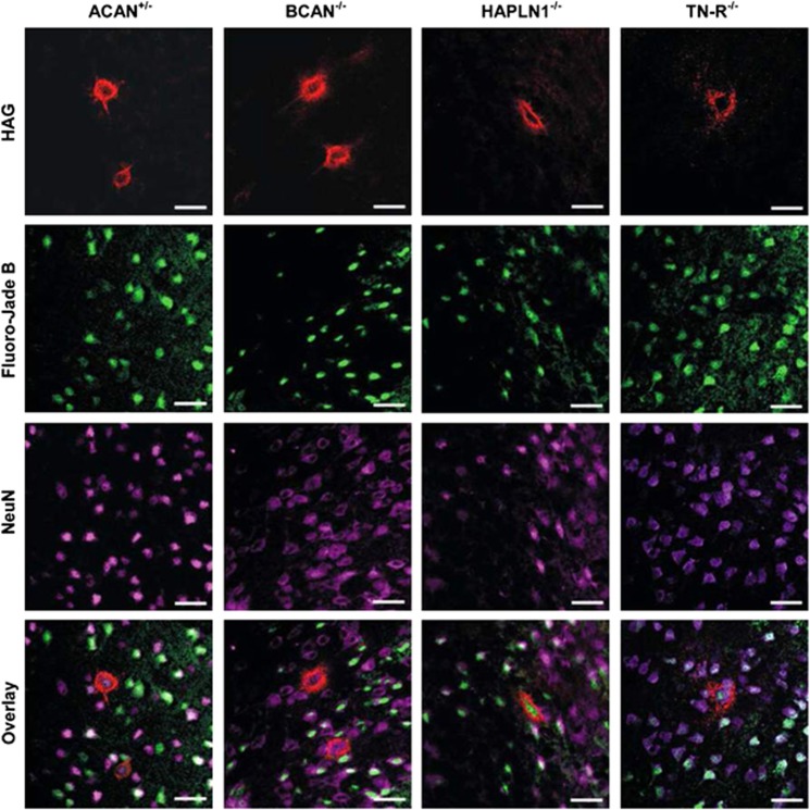
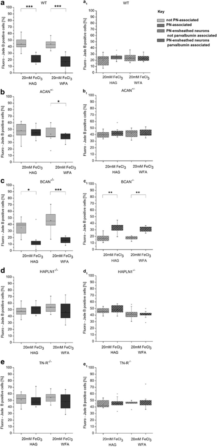
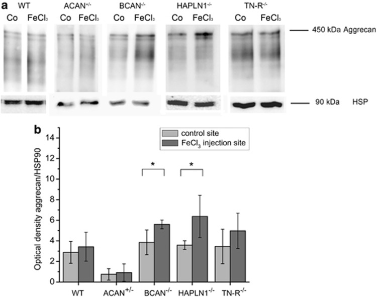
In Alzheimer's disease (AD), different types of neurons and different brain areas show differential patterns of vulnerability towards neurofibrillary degeneration, which provides the basis for a highly predictive profile of disease progression throughout the brain that now is widely accepted for neuropathological staging. In previous studies we could demonstrate that in AD cortical and subcortical neurons are constantly less frequently affected by neurofibrillary degeneration if they are enwrapped by a specialized form of the hyaluronan-based extracellular matrix (ECM), the so called 'perineuronal net' (PN). PNs are basically composed of large aggregating chondroitin sulphate proteoglycans connected to a hyaluronan backbone, stabilized by link proteins and cross-linked via tenascin-R (TN-R). Under experimental conditions in mice, PN-ensheathed neurons are better protected against iron-induced neurodegeneration than neurons without PN. Still, it remains unclear whether these neuroprotective effects are directly mediated by the PNs or are associated with some other mechanism in these neurons unrelated to PNs. To identify molecular components that essentially mediate the neuroprotective aspect on PN-ensheathed neurons, we comparatively analysed neuronal degeneration induced by a single injection of FeCl3 on four different mice knockout strains, each being deficient for a different component of PNs. Aggrecan, link protein and TN-R were identified to be essential for the neuroprotective properties of PN, whereas the contribution of brevican was negligible. Our findings indicate that the protection of PN-ensheathed neurons is directly mediated by the net structure and that both the high negative charge and the correct interaction of net components are essential for their neuroprotective function.
在阿尔茨海默病(AD)中,不同类型的神经元和不同脑区对神经原纤维变性表现出不同的易损模式,这为整个大脑疾病进展的高度预测性特征提供了基础,目前该特征已被广泛用于神经病理学分期。在先前的研究中,我们可以证明,在AD中,皮质和皮质下神经元如果被一种特殊形式的基于透明质酸的细胞外基质(ECM),即所谓的“神经周网”(PN)包裹,那么它们受神经原纤维变性影响的频率会持续较低。PN基本上由与透明质酸主链相连的大型聚集硫酸软骨素蛋白聚糖组成,通过连接蛋白稳定,并通过腱生蛋白-R(TN-R)交联。在小鼠的实验条件下,被PN包裹的神经元比没有PN的神经元更能抵抗铁诱导的神经变性。然而,这些神经保护作用是由PN直接介导的,还是与这些神经元中与PN无关的其他机制有关,仍不清楚。为了确定本质上介导对被PN包裹神经元神经保护作用的分子成分,我们比较分析了单次注射FeCl3在四种不同小鼠基因敲除品系中诱导的神经元变性,每个品系缺乏PN的不同成分。聚集蛋白聚糖、连接蛋白和TN-R被确定对PN的神经保护特性至关重要,而短蛋白聚糖的作用可忽略不计。我们的研究结果表明,对被PN包裹神经元的保护是由网络结构直接介导的,并且网络成分的高负电荷和正确相互作用对其神经保护功能至关重要。