Cui Xiaodong, Zhang Xiaoyun, Yin Qingling, Meng Aixia, Su Shaojuan, Jing Xu, Li Hong, Guan Xiumei, Li Xin, Liu Shunmei, Cheng Min
Medical Research Center, Weifang Medical University, Weifang, Shandong 261053, P.R. China.
Mol Med Rep. 2014 May;9(5):1641-7. doi: 10.3892/mmr.2014.2036. Epub 2014 Mar 11.
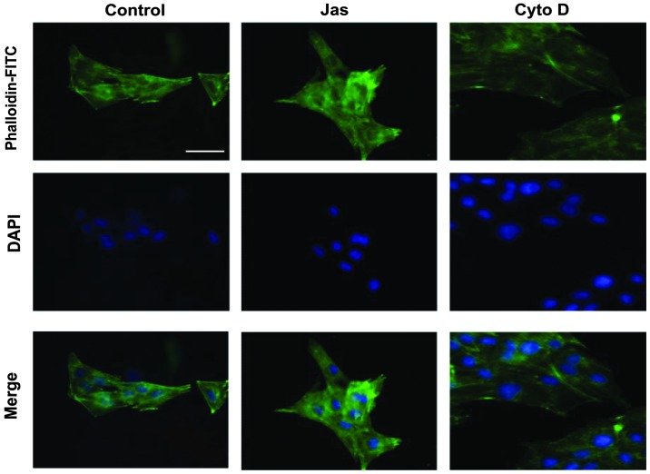
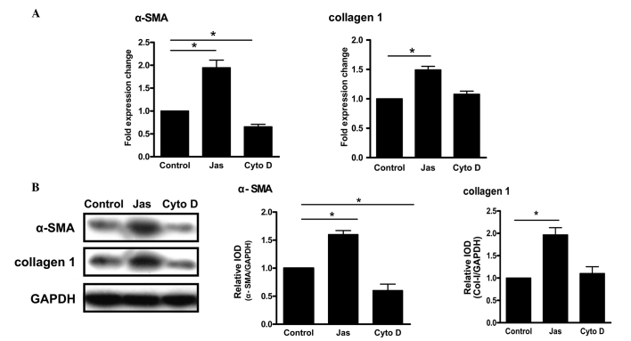
The activation of hepatic stellate cells (HSCs) is involved in the development of hepatic fibrosis. Previous studies have indicated that the acquisition of certain properties by activated HSCs is highly dependent on the reorganization of the actin cytoskeleton. However, direct evidence showing that the reorganization of the actin cytoskeleton is responsible for HSC activation is lacking. The aim of the present study was to investigate the role of cytoskeletal reorganization during HSC activation and to clarify the underlying mechanism. HSC-T6 cells were treated either with the F-actin stabilizer jasplakinolide (Jas) or the depolymerizer cytochalasin D (Cyto D). The actin cytoskeleton was evaluated via assessment of stress fiber formation. Furthermore, the activation properties of HSCs, including proliferation, adhesion, migration and the expression of α-smooth muscle actin (α-SMA) and collagen 1, were investigated in vitro. The results showed that Jas and Cyto D affected the actin distribution in HSC-T6 cells. Treatment with Jas resulted in thick actin bundles and a patchy appearance in the cytoplasm in HSC-T6 cells. In parallel, polymerization of actin microfilaments induced by Jas upregulated the expression of α-SMA and collagen 1, and also enhanced the migration and adhesion properties of HSC-T6 cells. Furthermore, the activation of HSC-T6 cells induced by the reorganization of the actin cytoskeleton was associated with the p38 mitogen-activated protein kinase (p38 MAPK) pathway. In conclusion, the present study suggests that the reorganization of the F-actin cytoskeleton is associated with HSC activation and that the p38 MAPK pathway is involved in this process. The inhibition of F-actin reorganization may thus be a potential key factor or molecular target for the control of liver fibrosis or cirrhosis.
肝星状细胞(HSCs)的激活参与肝纤维化的发展。先前的研究表明,活化的肝星状细胞获得某些特性高度依赖于肌动蛋白细胞骨架的重组。然而,缺乏直接证据表明肌动蛋白细胞骨架的重组是肝星状细胞激活的原因。本研究的目的是探讨细胞骨架重组在肝星状细胞激活过程中的作用,并阐明其潜在机制。用F-肌动蛋白稳定剂茉莉素内酯(Jas)或解聚剂细胞松弛素D(Cyto D)处理HSC-T6细胞。通过评估应力纤维形成来评价肌动蛋白细胞骨架。此外,在体外研究了肝星状细胞的激活特性,包括增殖、粘附、迁移以及α-平滑肌肌动蛋白(α-SMA)和胶原蛋白1的表达。结果表明,Jas和Cyto D影响HSC-T6细胞中的肌动蛋白分布。用Jas处理导致HSC-T6细胞中肌动蛋白束变粗且细胞质中出现斑点状外观。同时,Jas诱导的肌动蛋白微丝聚合上调了α-SMA和胶原蛋白1的表达,还增强了HSC-T6细胞的迁移和粘附特性。此外,肌动蛋白细胞骨架重组诱导的HSC-T6细胞激活与p38丝裂原活化蛋白激酶(p38 MAPK)途径有关。总之,本研究表明F-肌动蛋白细胞骨架的重组与肝星状细胞激活有关,并且p38 MAPK途径参与了这一过程。因此,抑制F-肌动蛋白重组可能是控制肝纤维化或肝硬化的潜在关键因素或分子靶点。