Hsieh Li-Chun, Chen John W, Wang Li-Ying, Tsang Yuk-Ming, Shueng Pei-Wei, Liao Li-Jen, Lo Wu-Chia, Lin Yu-Chin, Tseng Chien-Fu, Kuo Ying-Shiung, Jhuang Jie-Yang, Tien Hui-Ju, Juan Hsueh-Fen, Hsieh Chen-Hsi
Division of Medical Imaging, Department of Radiology, Far Eastern Memorial Hospital, Taipei, Taiwan; Department of Life Science, Institute of Molecular and Cellular Biology, Center for Systems Biology, National Taiwan University, Taipei, Taiwan; Medical Imaging Center, Taipei Medical University Hospital, Taipei, Taiwan.
Center for Systems Biology and Department of Radiology, Massachusetts General Hospital, Harvard Medical School, Massachusetts, United States of America.
PLoS One. 2014 Mar 21;9(3):e92561. doi: 10.1371/journal.pone.0092561. eCollection 2014.
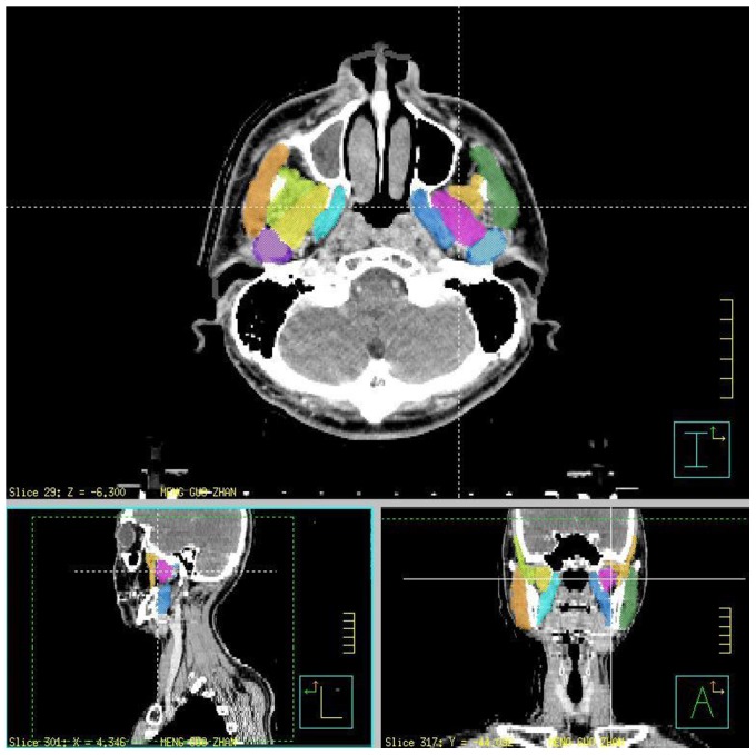
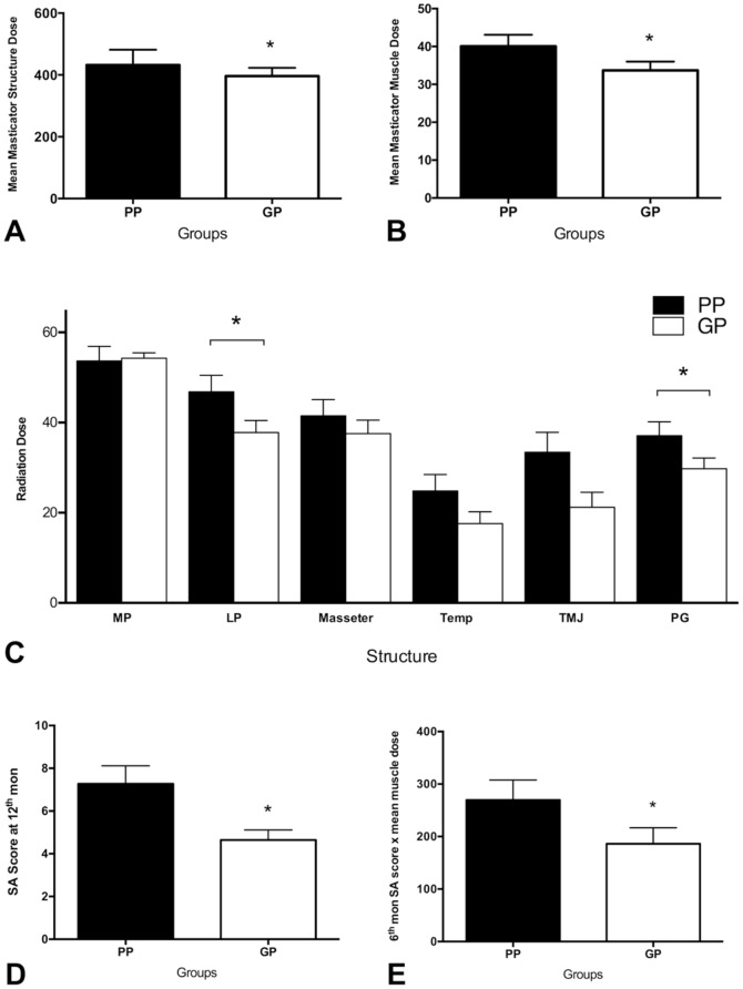
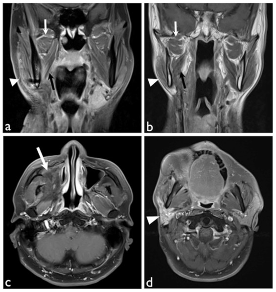
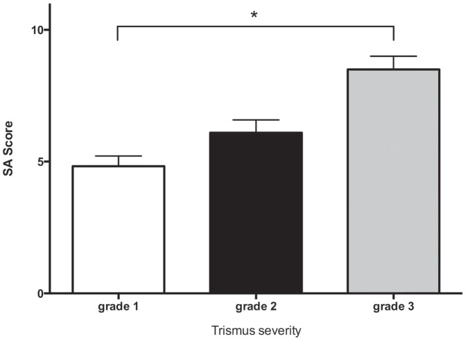
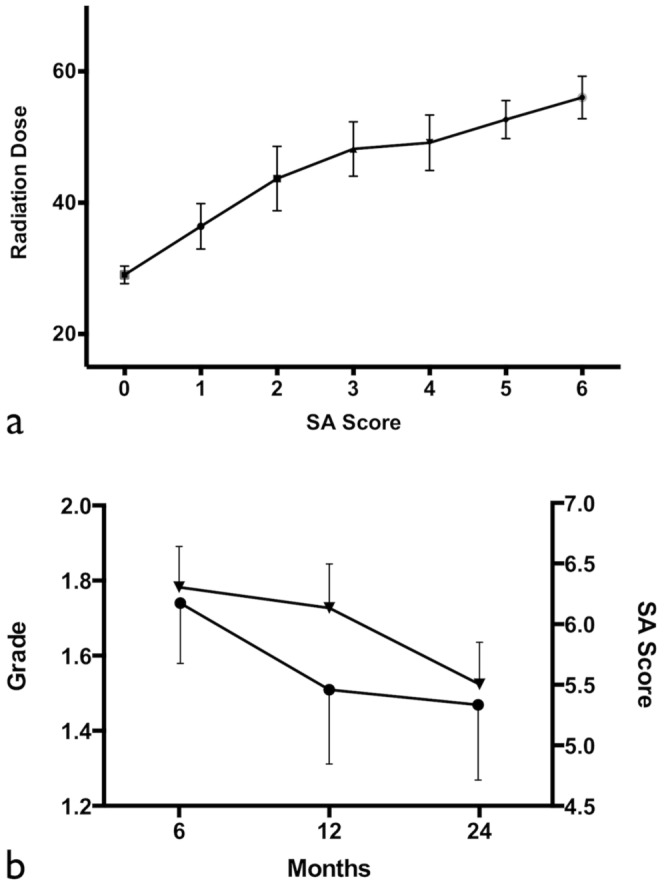
To develop magnetic resonance imaging (MRI) indicators to predict trismus outcome for post-operative oral cavity cancer patients who received adjuvant intensity-modulated radiation therapy (IMRT), 22 patients with oral cancer treated with IMRT were studied over a two-year period. Signal abnormality scores (SA scores) were computed from Likert-type ratings of the abnormalities of nine masticator structures and compared with the Mann-Whitney U-test and Kruskal-Wallis one-way ANOVA test between groups. Seventeen patients (77.3%) experienced different degrees of trismus during the two-year follow-up period. The SA score correlated with the trismus grade (r = 0.52, p<0.005). Patients having progressive trismus had higher mean doses of radiation to multiple structures, including the masticator and lateral pterygoid muscles, and the parotid gland (p<0.05). In addition, this group also had higher SA-masticator muscle dose product at 6 months and SA scores at 12 months (p<0.05). At the optimum cut-off points of 0.38 for the propensity score, the sensitivity was 100% and the specificity was 93% for predicting the prognosis of the trismus patients. The SA score, as determined using MRI, can reflect the radiation injury and correlate to trismus severity. Together with the radiation dose, it could serve as a useful biomarker to predict the outcome and guide the management of trismus following radiation therapy.
为了开发磁共振成像(MRI)指标,以预测接受辅助调强放射治疗(IMRT)的口腔癌术后患者的牙关紧闭结局,在两年时间里对22例接受IMRT治疗的口腔癌患者进行了研究。信号异常评分(SA评分)根据9个咀嚼肌结构异常的李克特式评分计算得出,并在组间采用曼-惠特尼U检验和克鲁斯卡尔-沃利斯单向方差分析进行比较。17例患者(77.3%)在两年随访期内出现不同程度的牙关紧闭。SA评分与牙关紧闭分级相关(r = 0.52,p<0.005)。牙关紧闭进展的患者对多个结构,包括咀嚼肌、翼外肌和腮腺,接受的平均辐射剂量更高(p<0.05)。此外,该组在6个月时的SA-咀嚼肌剂量乘积和12个月时的SA评分也更高(p<0.05)。在倾向评分为0.38的最佳截断点时,预测牙关紧闭患者预后的敏感性为100%,特异性为93%。MRI测定的SA评分可反映放射损伤并与牙关紧闭严重程度相关。结合放射剂量,它可作为预测放疗后牙关紧闭结局和指导其管理的有用生物标志物。