Sepehr Reyhaneh, Audi Said H, Maleki Sepideh, Staniszewski Kevin, Eis Annie L, Konduri Girija G, Ranji Mahsa
Biophotonics Laboratory, Department of Electrical Engineering and Computer Science, University of Wisconsin Milwaukee 3200 N Cramer St., Milwaukee, WI 53211, USA.
Department of Biomedical Engineering, Marquette University, 1515 W Wisconsin Avenue Milwaukee, WI 53233, USA.
J Innov Opt Health Sci. 2013 Jul 1;6(3):1350017. doi: 10.1142/S179354581350017X.
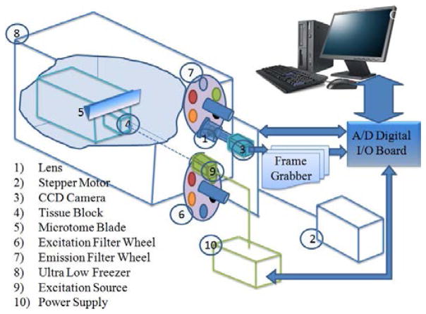
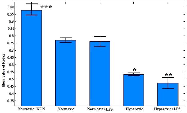
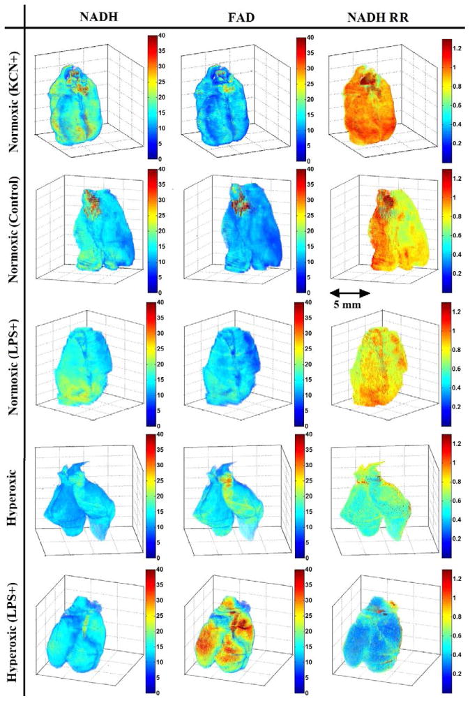
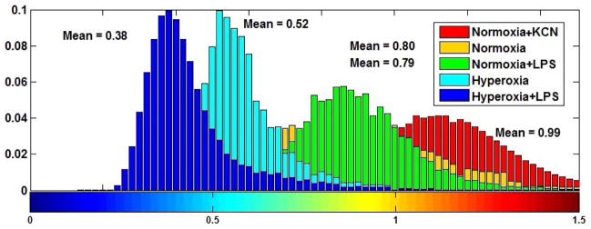
Reactive oxygen species (ROS) have been implicated in the pathogenesis of many acute and chronic pulmonary disorders such as acute lung injury (ALI) in adults and bronchopulmonary dysplasia (BPD) in premature infants. Bacterial infection and oxygen toxicity, which result in pulmonary vascular endothelial injury, contribute to impaired vascular growth and alveolar simplification seen in the lungs of premature infants with BPD. Hyperoxia induces ALI, reduces cell proliferation, causes DNA damage and promotes cell death by causing mitochondrial dysfunction. The objective of this study was to use an optical imaging technique to evaluate the variations in fluorescence intensities of the auto-fluorescent mitochondrial metabolic coenzymes, NADH and FAD in four different groups of rats. The ratio of these fluorescence signals (NADH/FAD), referred to as NADH redox ratio (NADH RR) has been used as an indicator of tissue metabolism in injuries. Here, we investigated whether the changes in metabolic state can be used as a marker of oxidative stress caused by hyperoxia and bacterial lipopolysaccharide (LPS) exposure in neonatal rat lungs. We examined the tissue redox states of lungs from four groups of rat pups: normoxic (21% O) pups, hyperoxic (90% O) pups, pups treated with LPS (normoxic + LPS), and pups treated with LPS and hyperoxia (hyperoxic + LPS). Our results show that hyperoxia oxidized the respiratory chain as reflected by a ~31% decrease in lung tissue NADH RR as compared to that for normoxic lungs. LPS treatment alone or with hyperoxia had no significant effect on lung tissue NADH RR as compared to that for normoxic or hyperoxic lungs, respectively. Thus, NADH RR serves as a quantitative marker of oxidative stress level in lung injury caused by two clinically important conditions: hyperoxia and LPS exposure.
活性氧(ROS)与许多急性和慢性肺部疾病的发病机制有关,如成人急性肺损伤(ALI)和早产儿支气管肺发育不良(BPD)。细菌感染和氧毒性会导致肺血管内皮损伤,这与患有BPD的早产儿肺部血管生长受损和肺泡简化有关。高氧会诱发ALI,减少细胞增殖,导致DNA损伤,并通过引起线粒体功能障碍促进细胞死亡。本研究的目的是使用光学成像技术评估四组不同大鼠中自荧光线粒体代谢辅酶NADH和FAD的荧光强度变化。这些荧光信号的比率(NADH/FAD),即NADH氧化还原比率(NADH RR),已被用作损伤中组织代谢的指标。在这里,我们研究了代谢状态的变化是否可以作为新生大鼠肺部高氧和细菌脂多糖(LPS)暴露引起的氧化应激的标志物。我们检查了四组幼鼠肺组织的氧化还原状态:常氧(21% O)幼鼠、高氧(90% O)幼鼠、LPS处理的幼鼠(常氧 + LPS)和LPS与高氧联合处理的幼鼠(高氧 + LPS)。我们的结果表明,与常氧肺相比,高氧使呼吸链氧化,肺组织NADH RR降低约31%。单独使用LPS或与高氧联合处理,与常氧或高氧肺相比,对肺组织NADH RR均无显著影响。因此,NADH RR可作为由两种临床重要情况引起的肺损伤中氧化应激水平的定量标志物:高氧和LPS暴露。