Cochain Clément, Chaudhari Sweena M, Koch Miriam, Wiendl Heinz, Eckstein Hans-Henning, Zernecke Alma
Institute of Clinical Biochemistry and Pathobiochemistry, University Hospital Würzburg, Würzburg, Germany; Department of Vascular Surgery, Klinikum rechts der Isar, Technical University Munich, Munich, Germany.
Institute of Clinical Biochemistry and Pathobiochemistry, University Hospital Würzburg, Würzburg, Germany.
PLoS One. 2014 Apr 1;9(4):e93280. doi: 10.1371/journal.pone.0093280. eCollection 2014.
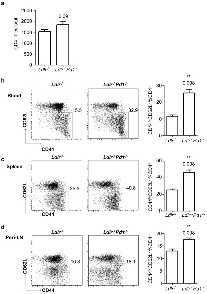
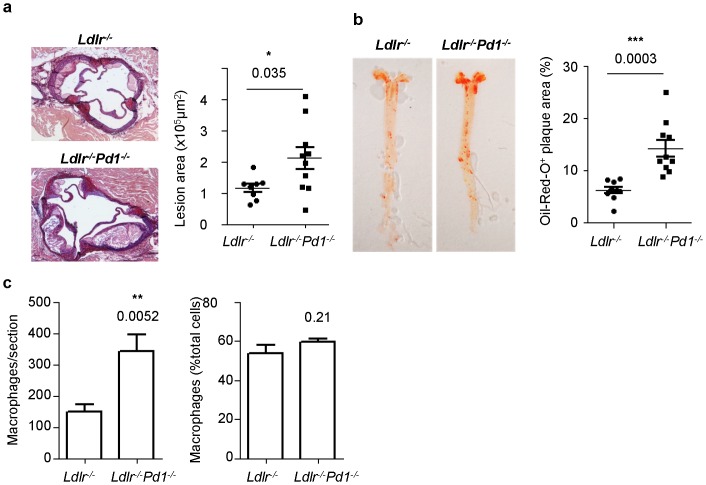
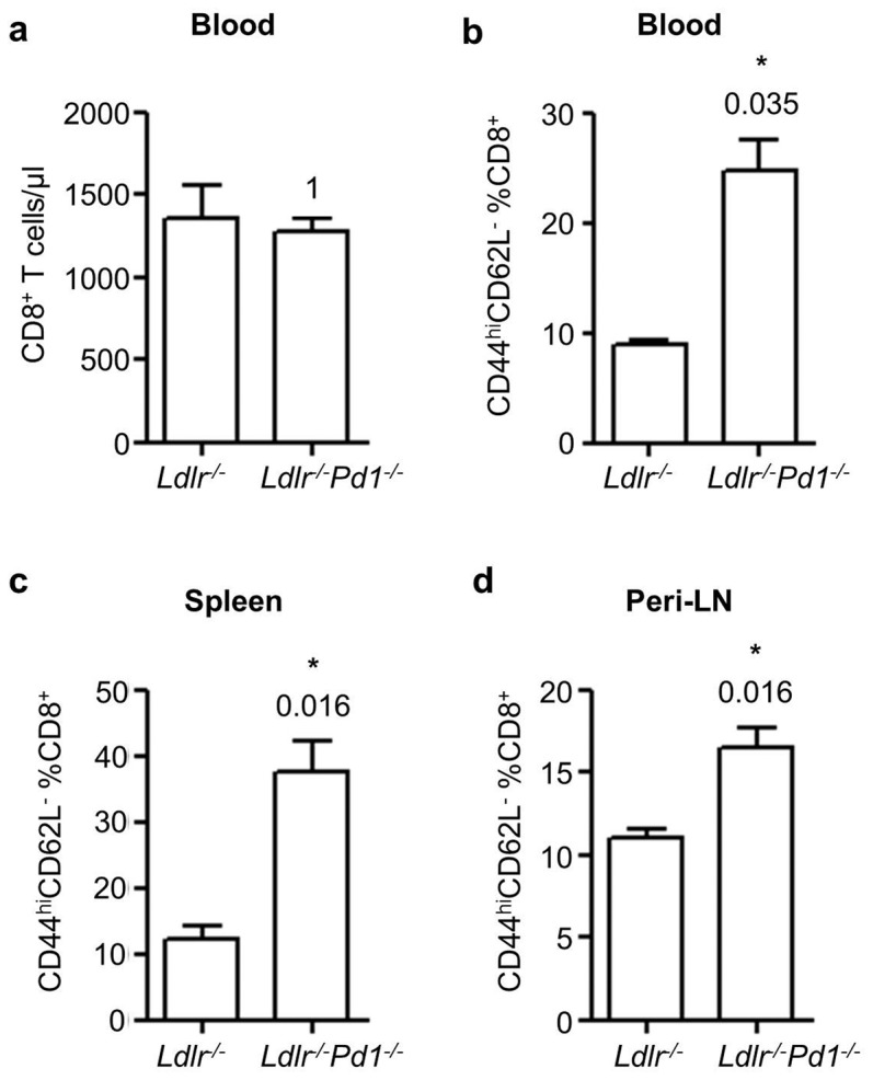
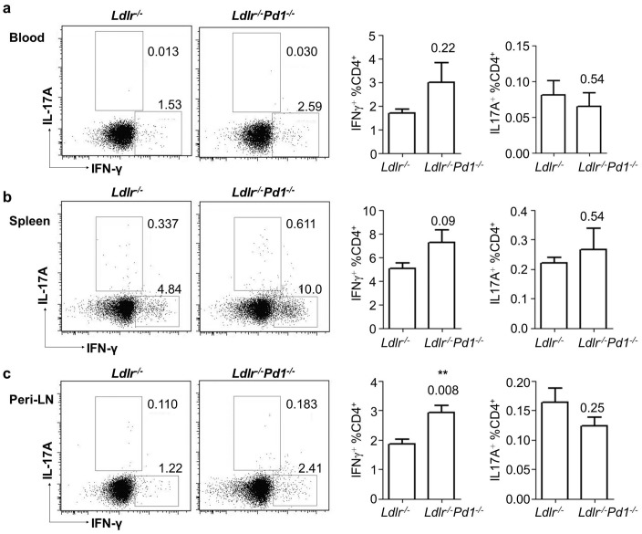
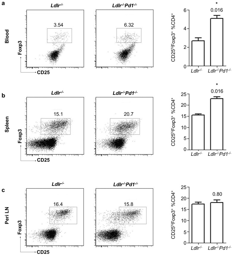
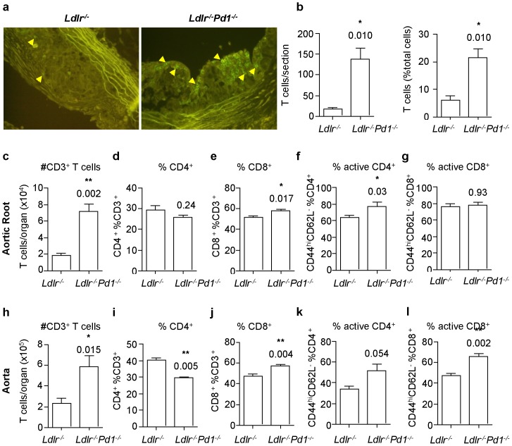
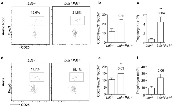
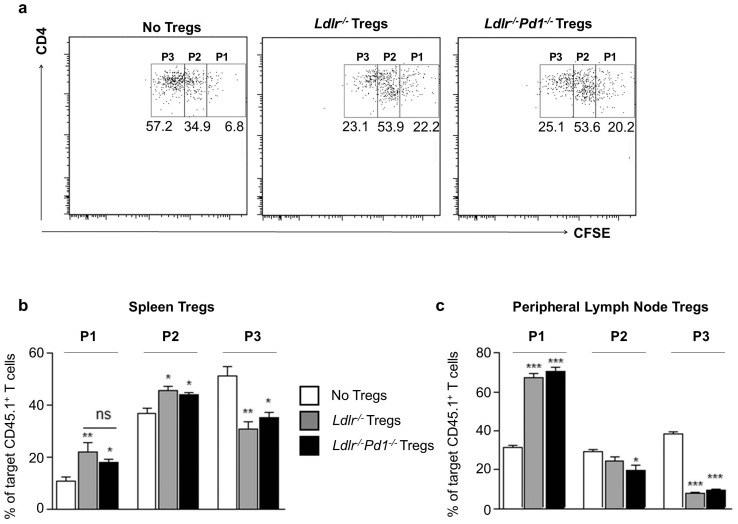
T cell activation represents a double-edged sword in atherogenesis, as it promotes both pro-inflammatory T cell activation and atheroprotective Foxp3(+) regulatory T cell (Treg) responses. Here, we investigated the role of the co-inhibitory receptor programmed cell death-1 (PD-1) in T cell activation and CD4(+) T cell polarization towards pro-atherogenic or atheroprotective responses in mice. Mice deficient for both low density lipoprotein receptor and PD-1 (Ldlr(-/-)Pd1(-/-)) displayed striking increases in systemic CD4(+) and CD8(+) T cell activation after 9 weeks of high fat diet feeding, associated with an expansion of both pro-atherogenic IFNγ-secreting T helper 1 cells and atheroprotective Foxp3+ Tregs. Importantly, PD-1 deficiency did not affect Treg suppressive function in vitro. Notably, PD-1 deficiency exacerbated atherosclerotic lesion growth and entailed a massive infiltration of T cells in atherosclerotic lesions. In addition, aggravated hypercholesterolemia was observed in Ldlr(-/-)Pd1(-/-) mice. In conclusion, we here demonstrate that although disruption of PD-1 signaling enhances both pro- and anti-atherogenic T cell responses in Ldlr(-/-) mice, pro-inflammatory T cell activation prevails and enhances dyslipidemia, vascular inflammation and atherosclerosis.
在动脉粥样硬化形成过程中,T细胞活化是一把双刃剑,因为它既促进促炎T细胞活化,也促进具有动脉粥样硬化保护作用的Foxp3(+)调节性T细胞(Treg)反应。在此,我们研究了共抑制受体程序性细胞死亡蛋白1(PD-1)在小鼠T细胞活化以及CD4(+) T细胞向促动脉粥样硬化或具有动脉粥样硬化保护作用的反应极化过程中的作用。低密度脂蛋白受体和PD-1均缺陷的小鼠(Ldlr(-/-)Pd1(-/-))在高脂饮食喂养9周后,全身CD4(+)和CD8(+) T细胞活化显著增加,这与促动脉粥样硬化的分泌IFNγ的辅助性T细胞1和具有动脉粥样硬化保护作用的Foxp3+ Tregs的扩增有关。重要的是,PD-1缺陷在体外并不影响Treg的抑制功能。值得注意的是,PD-1缺陷加剧了动脉粥样硬化病变的生长,并导致动脉粥样硬化病变中有大量T细胞浸润。此外,在Ldlr(-/-)Pd1(-/-)小鼠中观察到高胆固醇血症加重。总之,我们在此证明,尽管PD-1信号通路的破坏增强了Ldlr(-/-)小鼠中促动脉粥样硬化和抗动脉粥样硬化的T细胞反应,但促炎T细胞活化占主导并加剧了血脂异常、血管炎症和动脉粥样硬化。