• 文献检索
  • 文档翻译
  • 深度研究
  • 学术资讯
  • Suppr Zotero 插件Zotero 插件
  • 邀请有礼
  • 套餐&价格
  • 历史记录
应用&插件
Suppr Zotero 插件Zotero 插件浏览器插件Mac 客户端Windows 客户端微信小程序
定价
高级版会员购买积分包购买API积分包
服务
文献检索文档翻译深度研究API 文档MCP 服务
关于我们
关于 Suppr公司介绍联系我们用户协议隐私条款
关注我们

Suppr 超能文献

核心技术专利:CN118964589B侵权必究
粤ICP备2023148730 号-1Suppr @ 2026

文献检索

告别复杂PubMed语法,用中文像聊天一样搜索,搜遍4000万医学文献。AI智能推荐,让科研检索更轻松。

立即免费搜索

文件翻译

保留排版,准确专业,支持PDF/Word/PPT等文件格式,支持 12+语言互译。

免费翻译文档

深度研究

AI帮你快速写综述,25分钟生成高质量综述,智能提取关键信息,辅助科研写作。

立即免费体验

砷和铅对人脂肪来源间充质干细胞(AMSCs)的细胞毒性和遗传毒性作用。

Cytotoxic and Genotoxic effects of Arsenic and Lead on Human Adipose Derived Mesenchymal Stem Cells (AMSCs).

作者信息

Shakoori Ar, Ahmad A

机构信息

School of Biological Sciences, University of the Punjab , Quid-i-Azam Campus, Lahore, 54590. Pakistan.

出版信息

J Stem Cells Regen Med. 2013 Oct 15;9(2):29-36. doi: 10.46582/jsrm.0902007. eCollection 2013.

DOI:10.46582/jsrm.0902007
PMID:24693207
原文链接:https://pmc.ncbi.nlm.nih.gov/articles/PMC3908312/
Abstract

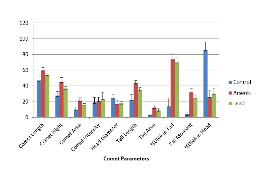
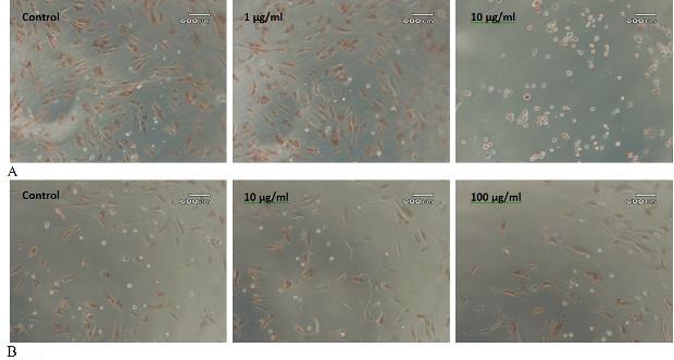
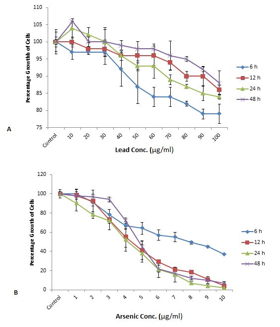
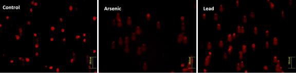
Arsenic and lead, known to have genotoxic and mutagenic effects, are ubiquitously distributed in the environment. The presence of arsenic in drinking water has been a serious health problem in many countries. Human exposure to these metals has also increased due to rapid industrialization and their use in formulation of many products. Liposuction material is a rich source of stem cells. In the present study cytotoxic and genotoxic effects of these metals were tested on adipose derived mesenchymal stem cells (AMSCs). Cells were exposed to 1-10 μg/ml and 10-100 μg/ml concentration of arsenic and lead, respectively, for 6, 12, 24 and 48 h. The cytotoxic effects were measured by neutral red uptake assay, while the genotoxic effects were tested by comet assay. The growth of cells decreased with increasing concentration and the duration of exposure to arsenic. Even the morphology of cells was changed; they became round at 10 μg /ml of arsenic. The cell growth was also decreased after exposure to lead, though it proved to be less toxic when cells were exposed for longer duration. The cell morphology remained unchanged. DNA damage was observed in the metal treated cells. Different parameters of comet assay were investigated for control and treated cells which indicated more DNA damage in arsenic treated cells compared to that of lead. Intact nuclei were observed in control cells. Present study clearly demonstrates that both arsenic and lead have cytotoxic and genotoxic effects on AMSCs, though arsenic compared to lead has more deleterious effects on AMSCs.

摘要

已知具有遗传毒性和致突变性的砷和铅在环境中广泛分布。饮用水中砷的存在在许多国家一直是严重的健康问题。由于快速工业化以及它们在许多产品配方中的使用,人类对这些金属的接触也有所增加。抽脂材料是干细胞的丰富来源。在本研究中,测试了这些金属对脂肪来源间充质干细胞(AMSCs)的细胞毒性和遗传毒性作用。细胞分别暴露于1 - 10μg/ml和10 - 100μg/ml浓度的砷和铅中,持续6、12、24和48小时。通过中性红摄取试验测量细胞毒性作用,同时通过彗星试验测试遗传毒性作用。随着砷浓度和暴露时间的增加,细胞生长减少。甚至细胞形态也发生了变化;在10μg/ml的砷浓度下它们变成了圆形。暴露于铅后细胞生长也减少,不过当细胞暴露较长时间时,铅的毒性较小。细胞形态保持不变。在金属处理的细胞中观察到DNA损伤。对对照细胞和处理细胞研究了彗星试验的不同参数,结果表明与铅处理的细胞相比,砷处理的细胞中DNA损伤更多。在对照细胞中观察到完整的细胞核。本研究清楚地表明,砷和铅对AMSCs都具有细胞毒性和遗传毒性作用,不过与铅相比,砷对AMSCs的有害影响更大。

https://cdn.ncbi.nlm.nih.gov/pmc/blobs/3a44/3908312/f7a91ccab571/jsrm-09-033-g005.jpg
https://cdn.ncbi.nlm.nih.gov/pmc/blobs/3a44/3908312/0ec67cc7f082/jsrm-09-031-g001.jpg
https://cdn.ncbi.nlm.nih.gov/pmc/blobs/3a44/3908312/638a7a95b029/jsrm-09-031-g002.jpg
https://cdn.ncbi.nlm.nih.gov/pmc/blobs/3a44/3908312/5a249a5be75a/jsrm-09-032-g003.jpg
https://cdn.ncbi.nlm.nih.gov/pmc/blobs/3a44/3908312/d23a626e3527/jsrm-09-033-g004.jpg
https://cdn.ncbi.nlm.nih.gov/pmc/blobs/3a44/3908312/f7a91ccab571/jsrm-09-033-g005.jpg

相似文献

1
Cytotoxic and Genotoxic effects of Arsenic and Lead on Human Adipose Derived Mesenchymal Stem Cells (AMSCs).砷和铅对人脂肪来源间充质干细胞(AMSCs)的细胞毒性和遗传毒性作用。
J Stem Cells Regen Med. 2013 Oct 15;9(2):29-36. doi: 10.46582/jsrm.0902007. eCollection 2013.
2
Comparative In Vitro Toxicity Evaluation of Heavy Metals (Lead, Cadmium, Arsenic, and Methylmercury) on HT-22 Hippocampal Cell Line.重金属(铅、镉、砷和甲基汞)对 HT-22 海马细胞系的体外比较毒性评价。
Biol Trace Elem Res. 2018 Jul;184(1):226-239. doi: 10.1007/s12011-017-1177-x. Epub 2017 Oct 9.
3
Uptake of inorganic and organic derivatives of arsenic associated with induced cytotoxic and genotoxic effects in Chinese hamster ovary (CHO) cells.砷的无机和有机衍生物的摄取与中国仓鼠卵巢(CHO)细胞中诱导的细胞毒性和基因毒性效应相关。
Toxicol Appl Pharmacol. 2004 Dec 1;201(2):156-65. doi: 10.1016/j.taap.2004.05.017.
4
Evaluation of genetic damage in tobacco and arsenic exposed population of Southern Assam, India using buccal cytome assay and comet assay.采用口腔细胞微核检测法和彗星检测法评估印度阿萨姆邦南部地区暴露于烟草和砷环境人群的遗传损伤。
Ecotoxicol Environ Saf. 2016 Feb;124:169-176. doi: 10.1016/j.ecoenv.2015.10.019. Epub 2015 Oct 27.
5
The effects of arsenic trioxide on DNA synthesis and genotoxicity in human colon cancer cells.三氧化二砷对人结肠癌细胞 DNA 合成和遗传毒性的影响。
Int J Environ Res Public Health. 2010 May;7(5):2018-32. doi: 10.3390/ijerph7052018. Epub 2010 Apr 28.
6
Hazards of heavy metal contamination.重金属污染的危害。
Br Med Bull. 2003;68:167-82. doi: 10.1093/bmb/ldg032.
7
Relative sensitivity of fish and mammalian cells to the antibiotic, trimethoprim: cytotoxic and genotoxic responses as determined by neutral red retention, Comet and micronucleus assays.鱼和哺乳动物细胞对抗生素甲氧苄啶的相对敏感性:通过中性红保留试验、彗星试验和微核试验测定的细胞毒性和遗传毒性反应。
Ecotoxicology. 2011 Jan;20(1):208-17. doi: 10.1007/s10646-010-0572-2. Epub 2010 Nov 21.
8
Investigation on the genotoxic effects of long-term administration of sodium arsenite in bone marrow and testicular cells in vivo using the comet assay.利用彗星试验对亚砷酸钠长期体内给药在骨髓和睾丸细胞中的遗传毒性作用进行研究。
J Environ Pathol Toxicol Oncol. 2007;26(1):29-37. doi: 10.1615/jenvironpatholtoxicoloncol.v26.i1.40.
9
Enhanced renoprotective effect of GDNF-modified adipose-derived mesenchymal stem cells on renal interstitial fibrosis.胶质细胞源性神经营养因子修饰的脂肪间充质干细胞对肾间质纤维化的增强肾脏保护作用。
Stem Cell Res Ther. 2021 Jan 7;12(1):27. doi: 10.1186/s13287-020-02049-z.
10
In-vitro cytotoxic and genotoxic effects of arsenic trioxide on human leukemia (HL-60) cells using the MTT and alkaline single cell gel electrophoresis (Comet) assays.采用MTT法和碱性单细胞凝胶电泳(彗星试验)检测三氧化二砷对人白血病(HL - 60)细胞的体外细胞毒性和遗传毒性作用。
Mol Cell Biochem. 2007 Jul;301(1-2):123-30. doi: 10.1007/s11010-006-9403-4. Epub 2007 Jan 10.

引用本文的文献

1
Assessing Acute and Chronic Risks of Human Exposure to Arsenic: A Cross-Sectional Study in Ethiopia Employing Body Biomarkers.评估人类接触砷的急性和慢性风险:埃塞俄比亚一项采用人体生物标志物的横断面研究。
Environ Health Insights. 2024 May 30;18:11786302241257365. doi: 10.1177/11786302241257365. eCollection 2024.
2
Examining carcinogenic and noncarcinogenic health risks related to arsenic exposure in Ethiopia: A longitudinal study.埃塞俄比亚砷暴露相关致癌和非致癌健康风险研究:一项纵向研究。
Toxicol Rep. 2024 Jan 3;12:100-110. doi: 10.1016/j.toxrep.2024.01.001. eCollection 2024 Jun.
3
A Comprehensive Overview of the Complex Role of Oxidative Stress in Aging, The Contributing Environmental Stressors and Emerging Antioxidant Therapeutic Interventions.

本文引用的文献

1
Investigating the effect of lead acetate on rat bone marrow-derived mesenchymal stem cells toxicity: role of apoptosis.探讨醋酸铅对大鼠骨髓间充质干细胞毒性的影响:凋亡的作用。
Toxicol Mech Methods. 2011 Mar;21(3):225-30. doi: 10.3109/15376516.2010.543943. Epub 2011 Jan 18.
2
Arsenite induces apoptosis in human mesenchymal stem cells by altering Bcl-2 family proteins and by activating intrinsic pathway.亚砷酸盐通过改变 Bcl-2 家族蛋白和激活内在途径诱导人骨髓间充质干细胞凋亡。
Toxicol Appl Pharmacol. 2010 May 1;244(3):263-72. doi: 10.1016/j.taap.2010.01.001. Epub 2010 Jan 18.
3
Arsenic trioxide inhibits growth of As4.1 juxtaglomerular cells via cell cycle arrest and caspase-independent apoptosis.
氧化应激在衰老中的复杂作用、相关环境应激源及新兴抗氧化治疗干预的综合概述
Front Aging Neurosci. 2022 Jun 13;14:827900. doi: 10.3389/fnagi.2022.827900. eCollection 2022.
4
Effects of Oral Dosage of Lead Acetate II on Osteocalcin Gene Expression in Rat Mesenchymal Stem Cells.口服醋酸铅II对大鼠间充质干细胞骨钙素基因表达的影响
Rep Biochem Mol Biol. 2018 Apr;6(2):131-136.
5
Stem cell research in pakistan; past, present and future.巴基斯坦的干细胞研究:过去、现在与未来。
Int J Stem Cells. 2015 May;8(1):1-8. doi: 10.15283/ijsc.2015.8.1.1.
三氧化二砷通过细胞周期阻滞和非半胱天冬酶依赖性凋亡抑制As4.1肾小球旁细胞的生长。
Am J Physiol Renal Physiol. 2007 Aug;293(2):F511-20. doi: 10.1152/ajprenal.00385.2006. Epub 2007 May 16.
4
Cyclin B1 proteolysis via p38 MAPK signaling participates in G2 checkpoint elicited by arsenite.通过p38丝裂原活化蛋白激酶信号传导的细胞周期蛋白B1蛋白水解参与亚砷酸盐引发的G2期检查点。
J Cell Physiol. 2007 Aug;212(2):481-8. doi: 10.1002/jcp.21042.
5
Transplacental arsenic carcinogenesis in mice.小鼠经胎盘的砷致癌作用。
Toxicol Appl Pharmacol. 2007 Aug 1;222(3):271-80. doi: 10.1016/j.taap.2006.12.034. Epub 2007 Jan 12.
6
Arsenic exposure is associated with decreased DNA repair in vitro and in individuals exposed to drinking water arsenic.砷暴露与体外以及饮用水砷暴露个体的DNA修复能力下降有关。
Environ Health Perspect. 2006 Aug;114(8):1193-8. doi: 10.1289/ehp.9008.
7
Adipose tissue-derived mesenchymal stem cell yield and growth characteristics are affected by the tissue-harvesting procedure.脂肪组织来源的间充质干细胞产量和生长特性受组织采集程序的影响。
Cytotherapy. 2006;8(2):166-77. doi: 10.1080/14653240600621125.
8
Combination treatment with arsenic trioxide and sulindac augments their apoptotic potential in lung cancer cells through activation of caspase cascade and mitochondrial dysfunction.三氧化二砷与舒林酸联合治疗通过激活半胱天冬酶级联反应和线粒体功能障碍增强其对肺癌细胞的凋亡诱导潜力。
Int J Oncol. 2006 Jun;28(6):1401-8.
9
Lead toxicity, a review of the literature. Part 1: Exposure, evaluation, and treatment.铅中毒:文献综述。第1部分:暴露、评估与治疗。
Altern Med Rev. 2006 Mar;11(1):2-22.
10
Characterization of freshly isolated and cultured cells derived from the fatty and fluid portions of liposuction aspirates.对从抽脂吸出物的脂肪和液体部分分离出的新鲜细胞及培养细胞的特性分析。
J Cell Physiol. 2006 Jul;208(1):64-76. doi: 10.1002/jcp.20636.