• 文献检索
  • 文档翻译
  • 深度研究
  • 学术资讯
  • Suppr Zotero 插件Zotero 插件
  • 邀请有礼
  • 套餐&价格
  • 历史记录
应用&插件
Suppr Zotero 插件Zotero 插件浏览器插件Mac 客户端Windows 客户端微信小程序
定价
高级版会员购买积分包购买API积分包
服务
文献检索文档翻译深度研究API 文档MCP 服务
关于我们
关于 Suppr公司介绍联系我们用户协议隐私条款
关注我们

Suppr 超能文献

核心技术专利:CN118964589B侵权必究
粤ICP备2023148730 号-1Suppr @ 2026

文献检索

告别复杂PubMed语法,用中文像聊天一样搜索,搜遍4000万医学文献。AI智能推荐,让科研检索更轻松。

立即免费搜索

文件翻译

保留排版,准确专业,支持PDF/Word/PPT等文件格式,支持 12+语言互译。

免费翻译文档

深度研究

AI帮你快速写综述,25分钟生成高质量综述,智能提取关键信息,辅助科研写作。

立即免费体验

弥散加权磁共振成像用于弥漫性大B细胞淋巴瘤的分期及疗效评估:一项初步研究

Diffusion-weighted MRI for staging and evaluation of response in diffuse large B-cell lymphoma: a pilot study.

作者信息

Siegel Marilyn J, Jokerst Clint E, Rajderkar Dhana, Hildebolt Charles F, Goyal Sagun, Dehdashti Farrokh, Wagner Johnston Nina, Siegel Barry A

机构信息

Mallinckrodt Institute of Radiology, Washington University School of Medicine, St. Louis, MO, USA; Siteman Cancer Center, Washington University School of Medicine, St. Louis, MO, USA.

出版信息

NMR Biomed. 2014 Jun;27(6):681-91. doi: 10.1002/nbm.3105. Epub 2014 Apr 3.

DOI:10.1002/nbm.3105
PMID:24700565
原文链接:https://pmc.ncbi.nlm.nih.gov/articles/PMC4026031/
Abstract

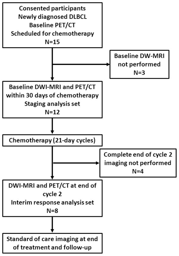
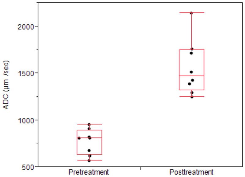
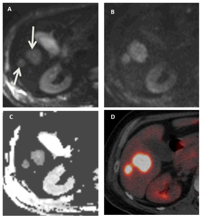
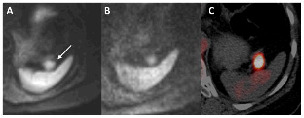
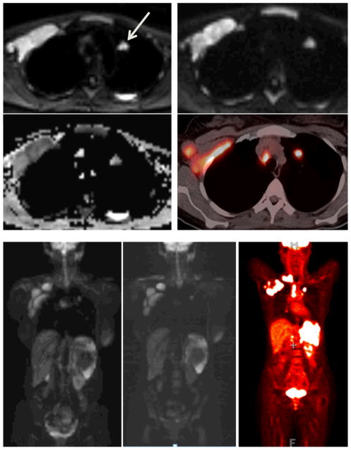
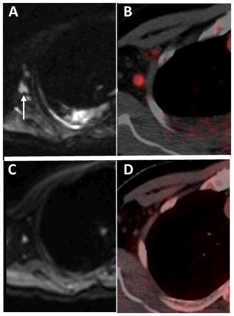
The aim of this study was to compare diffusion-weighted MRI (DW-MRI) with positron emission tomography/computed tomography (PET/CT) for the staging and evaluation of the treatment response in patients with diffuse large B-cell lymphoma (DLBCL). Institutional review board approval was obtained for this study; all subjects gave informed consent. Twelve patients were imaged before treatment and eight of these were also imaged after two cycles of chemotherapy using both DW-MRI and PET/CT. Up to six target lesions were selected at baseline for response assessment based on International Working Group criteria (nodes > 1.5 cm in diameter; extranodal lesions > 1 cm in diameter). For pretreatment staging, visual analysis of the numbers of nodal and extranodal lesions based on PET/CT was performed. For interim response assessment after cycle 2 of chemotherapy, residual tumor sites were assessed visually and the percentage changes in target lesion size, maximum standardized uptake value (SUVmax ) and apparent diffusion coefficient (ADC) from pretreatment values were calculated. In 12 patients studied pretreatment, there were 46 nodal and 16 extranodal sites of lymphomatous involvement. Agreement between DW-MRI and PET/CT for overall lesion detection was 97% (60/62 tumor sites; 44/46 nodal and 16/16 extranodal lesions) and, for Ann Arbor stage, it was 100%. In the eight patients who had interim assessment, five of their 49 tumor sites remained abnormal on visual analysis of both DW-MRI and PET/CT, and there was one false positive on DW-MRI. Of their 24 target lesions, the mean pretreatment ADC value, tumor size and SUVmax were 772 µm(2) /s, 21.3 cm(2) and 16.9 g/mL, respectively. At interim assessment of the same 24 target lesions, ADC values increased by 85%, tumor size decreased by 74% and SUVmax decreased by 83% (all p < 0.01 versus baseline). DW-MRI provides results comparable with those of PET/CT for staging and early response assessment in patients with DLBCL.

摘要

本研究旨在比较弥散加权磁共振成像(DW-MRI)与正电子发射断层扫描/计算机断层扫描(PET/CT)在弥漫性大B细胞淋巴瘤(DLBCL)患者分期及治疗反应评估中的应用。本研究获得了机构审查委员会的批准;所有受试者均签署了知情同意书。12例患者在治疗前进行了成像检查,其中8例在接受两个周期化疗后同时使用DW-MRI和PET/CT进行了成像检查。根据国际工作组标准(直径>1.5 cm的淋巴结;直径>1 cm的结外病变),在基线时选择多达6个靶病变进行反应评估。对于治疗前分期,基于PET/CT对淋巴结和结外病变的数量进行视觉分析。对于化疗第2周期后的中期反应评估,对残留肿瘤部位进行视觉评估,并计算靶病变大小、最大标准化摄取值(SUVmax)和表观扩散系数(ADC)相对于治疗前值的百分比变化。在12例接受治疗前研究的患者中,有46个淋巴结和16个结外部位存在淋巴瘤累及。DW-MRI与PET/CT在总体病变检测方面的一致性为97%(62个肿瘤部位中的60个;46个淋巴结中的44个和16个结外病变中的16个),在Ann Arbor分期方面,一致性为100%。在8例进行中期评估的患者中,在对DW-MRI和PET/CT的视觉分析中,其49个肿瘤部位中有5个仍异常,且DW-MRI有1例假阳性。在其24个靶病变中,治疗前ADC值、肿瘤大小和SUVmax的平均值分别为772 µm(2)/s、21.3 cm(2)和16.9 g/mL。在对相同的24个靶病变进行中期评估时,ADC值增加了85%,肿瘤大小减少了74%,SUVmax减少了83%(与基线相比,所有p<0.01)。DW-MRI在DLBCL患者的分期和早期反应评估中提供了与PET/CT相当的结果。

https://cdn.ncbi.nlm.nih.gov/pmc/blobs/ee38/4026031/b20cc34609fa/nihms577581f6.jpg
https://cdn.ncbi.nlm.nih.gov/pmc/blobs/ee38/4026031/7d92e35c54bd/nihms577581f1.jpg
https://cdn.ncbi.nlm.nih.gov/pmc/blobs/ee38/4026031/3b8a0c3c0c43/nihms577581f2.jpg
https://cdn.ncbi.nlm.nih.gov/pmc/blobs/ee38/4026031/8b7216336336/nihms577581f3.jpg
https://cdn.ncbi.nlm.nih.gov/pmc/blobs/ee38/4026031/2147fb911d5f/nihms577581f4a.jpg
https://cdn.ncbi.nlm.nih.gov/pmc/blobs/ee38/4026031/f443920b4f5c/nihms577581f5.jpg
https://cdn.ncbi.nlm.nih.gov/pmc/blobs/ee38/4026031/b20cc34609fa/nihms577581f6.jpg

相似文献

1
Diffusion-weighted MRI for staging and evaluation of response in diffuse large B-cell lymphoma: a pilot study.弥散加权磁共振成像用于弥漫性大B细胞淋巴瘤的分期及疗效评估:一项初步研究
NMR Biomed. 2014 Jun;27(6):681-91. doi: 10.1002/nbm.3105. Epub 2014 Apr 3.
2
Diffusion-weighted MRI in early chemotherapy response evaluation of patients with diffuse large B-cell lymphoma--a pilot study: comparison with 2-deoxy-2-fluoro- D-glucose-positron emission tomography/computed tomography.弥散加权 MRI 在弥漫性大 B 细胞淋巴瘤患者早期化疗反应评估中的应用——一项初步研究:与 2-脱氧-2-氟-D-葡萄糖正电子发射断层扫描/计算机断层扫描的比较。
NMR Biomed. 2011 Dec;24(10):1181-90. doi: 10.1002/nbm.1689. Epub 2011 Mar 8.
3
Comparison of PET/MRI With PET/CT in the Evaluation of Disease Status in Lymphoma.PET/MRI与PET/CT在淋巴瘤疾病状态评估中的比较
Clin Nucl Med. 2017 Jan;42(1):e1-e7. doi: 10.1097/RLU.0000000000001344.
4
Whole-Body Diffusion-Weighted MRI Compared to FDG PET/CT in Initial Staging and Therapy Response Assessment of Hodgkin Lymphoma in Pediatric Patients.全身扩散加权 MRI 与 FDG PET/CT 在儿科霍奇金淋巴瘤初始分期和治疗反应评估中的比较。
Can Assoc Radiol J. 2020 May;71(2):217-225. doi: 10.1177/0846537119888380. Epub 2020 Jan 22.
5
[Informativeness of whole-body diffusion-weighted magnetic resonance imaging and positron emission tomography with computed tomography in follicular lymphoma].[全身弥散加权磁共振成像及正电子发射断层显像/X线计算机体层成像在滤泡性淋巴瘤中的诊断价值]
Ter Arkh. 2020 Sep 1;92(7):55-62. doi: 10.26442/00403660.2020.07.000774.
6
No correlation between glucose metabolism and apparent diffusion coefficient in diffuse large B-cell lymphoma: a PET/CT and DW-MRI study.弥漫性大 B 细胞淋巴瘤中葡萄糖代谢与表观扩散系数之间无相关性:一项 PET/CT 和 DW-MRI 研究。
Eur J Radiol. 2011 Aug;79(2):e117-21. doi: 10.1016/j.ejrad.2011.04.062. Epub 2011 May 18.
7
Diffusion-weighted MRI compared to FDG PET/CT for assessment of early treatment response in lymphoma.扩散加权磁共振成像与氟代脱氧葡萄糖正电子发射断层扫描/计算机断层扫描在评估淋巴瘤早期治疗反应中的比较
Acta Radiol. 2015 Feb;56(2):152-8. doi: 10.1177/0284185114526087. Epub 2014 Feb 28.
8
PET/MRI for the evaluation of patients with lymphoma: initial observations.用于淋巴瘤患者评估的PET/MRI:初步观察结果
AJR Am J Roentgenol. 2015 Apr;204(4):842-8. doi: 10.2214/AJR.14.13181.
9
Diagnostic performance of FDG-PET/MRI and WB-DW-MRI in the evaluation of lymphoma: a prospective comparison to standard FDG-PET/CT.18F-氟代脱氧葡萄糖正电子发射断层扫描/磁共振成像(FDG-PET/MRI)与全身扩散加权磁共振成像(WB-DW-MRI)在淋巴瘤评估中的诊断效能:与标准FDG-PET/CT的前瞻性比较
BMC Cancer. 2015 Dec 23;15:1002. doi: 10.1186/s12885-015-2009-z.
10
Standardized uptake value for (18)F-fluorodeoxyglucose is correlated with a high International Prognostic Index and the presence of extranodal involvement in patients with diffuse large B-cell lymphoma.(18)F-氟脱氧葡萄糖的标准化摄取值与弥漫性大B细胞淋巴瘤患者的高国际预后指数及结外受累情况相关。
Rev Esp Med Nucl Imagen Mol. 2014 May-Jun;33(3):148-52. doi: 10.1016/j.remn.2013.07.008. Epub 2013 Oct 2.

引用本文的文献

1
Whole-body magnetic resonance imaging for cutaneous melanoma staging: A scientific review.用于皮肤黑色素瘤分期的全身磁共振成像:一项科学综述。
World J Clin Oncol. 2025 Aug 24;16(8):109206. doi: 10.5306/wjco.v16.i8.109206.
2
Comparison of diffusion-weighted MRI and [F]FDG PET/MRI for treatment monitoring in pediatric Hodgkin and non-Hodgkin lymphoma.弥散加权 MRI 与 [F]FDG PET/MRI 对比在儿科霍奇金和非霍奇金淋巴瘤治疗监测中的应用。
Eur Radiol. 2024 Jan;34(1):643-653. doi: 10.1007/s00330-023-10015-5. Epub 2023 Aug 5.
3
Detection of bone marrow metastases in children and young adults with solid cancers with diffusion-weighted MRI.

本文引用的文献

1
Competing Technology for PET/Computed Tomography: Diffusion-weighted Magnetic Resonance Imaging.正电子发射断层扫描/计算机断层扫描的竞争技术:扩散加权磁共振成像
PET Clin. 2013 Jul;8(3):259-77. doi: 10.1016/j.cpet.2013.03.002. Epub 2013 May 17.
2
An international confirmatory study of the prognostic value of early PET/CT in diffuse large B-cell lymphoma: comparison between Deauville criteria and ΔSUVmax.一项早期 PET/CT 对弥漫性大 B 细胞淋巴瘤预后价值的国际确证性研究:Deauville 标准与 ΔSUVmax 的比较。
Eur J Nucl Med Mol Imaging. 2013 Sep;40(9):1312-20. doi: 10.1007/s00259-013-2435-6. Epub 2013 May 7.
3
Whole-body diffusion-weighted MRI: tips, tricks, and pitfalls.
应用弥散加权 MRI 检测儿童和青年实体瘤患者骨髓转移。
Skeletal Radiol. 2023 Jun;52(6):1179-1192. doi: 10.1007/s00256-022-04240-0. Epub 2022 Nov 28.
4
Whole-Body MRI Is an Effective Imaging Modality for Hematological Malignancy Treatment Response Assessment: A Systematic Review and Meta-Analysis.全身磁共振成像(MRI)是评估血液系统恶性肿瘤治疗反应的有效成像方式:一项系统评价和荟萃分析
Front Oncol. 2022 Feb 18;12:827777. doi: 10.3389/fonc.2022.827777. eCollection 2022.
5
Whole-body diffusion-weighted MRI in lymphoma-comparison of global apparent diffusion coefficient histogram parameters for differentiation of diseased nodes of lymphoma patients from normal lymph nodes of healthy individuals.淋巴瘤的全身扩散加权磁共振成像——比较淋巴瘤患者病变淋巴结与健康个体正常淋巴结的整体表观扩散系数直方图参数以进行鉴别
Quant Imaging Med Surg. 2021 Aug;11(8):3549-3561. doi: 10.21037/qims-21-50.
6
One-stop local and whole-body staging of children with cancer.一站式局部和全身分期诊断儿童癌症。
Pediatr Radiol. 2022 Feb;52(2):391-400. doi: 10.1007/s00247-021-05076-x. Epub 2021 Apr 30.
7
Therapy Response Assessment of Pediatric Tumors with Whole-Body Diffusion-weighted MRI and FDG PET/MRI.全身扩散加权 MRI 和 FDG PET/MRI 评估儿科肿瘤的治疗反应。
Radiology. 2020 Jul;296(1):143-151. doi: 10.1148/radiol.2020192508. Epub 2020 May 5.
8
Semiautomated Workflow for Clinically Streamlined Glioma Parametric Response Mapping.用于临床简化胶质瘤参数反应映射的半自动工作流程。
Tomography. 2016 Dec;2(4):267-275. doi: 10.18383/j.tom.2016.00181.
9
Osteonecrosis detected by whole body magnetic resonance in patients with Hodgkin Lymphoma treated by BEACOPP.采用BEACOPP方案治疗的霍奇金淋巴瘤患者中通过全身磁共振检测到的骨坏死
Eur Radiol. 2017 May;27(5):2129-2136. doi: 10.1007/s00330-016-4535-8. Epub 2016 Aug 13.
10
Intravoxel incoherent motion diffusion-weighted imaging for monitoring chemotherapeutic efficacy in gastric cancer.体素内不相干运动扩散加权成像用于监测胃癌化疗疗效
World J Gastroenterol. 2016 Jun 28;22(24):5520-31. doi: 10.3748/wjg.v22.i24.5520.
全身扩散加权磁共振成像:技巧、窍门和陷阱。
AJR Am J Roentgenol. 2012 Aug;199(2):252-62. doi: 10.2214/AJR.11.7866.
4
Combined modality treatment for PET-positive non-Hodgkin lymphoma: favorable outcomes of combined modality treatment for patients with non-Hodgkin lymphoma and positive interim or postchemotherapy FDG-PET.正电子发射断层扫描(PET)阳性非霍奇金淋巴瘤的联合治疗模式:伴有非霍奇金淋巴瘤且化疗后正电子发射断层扫描(PET)氟脱氧葡萄糖(FDG-PET)阳性的患者,采用联合治疗模式可获得良好的转归。
Int J Radiat Oncol Biol Phys. 2012 Aug 1;83(5):e647-54. doi: 10.1016/j.ijrobp.2012.01.060. Epub 2012 May 18.
5
Whole-body diffusion-weighted MR imaging in cancer: current status and research directions.全身扩散加权磁共振成像在癌症中的应用:现状与研究方向。
Radiology. 2011 Dec;261(3):700-18. doi: 10.1148/radiol.11110474.
6
Principles and applications of diffusion-weighted imaging in cancer detection, staging, and treatment follow-up.扩散加权成像在癌症检测、分期和治疗随访中的原理及应用。
Radiographics. 2011 Oct;31(6):1773-91. doi: 10.1148/rg.316115515.
7
PET in T-Cell Lymphoma.正电子发射断层扫描(PET)在 T 细胞淋巴瘤中的应用。
Curr Hematol Malig Rep. 2011 Dec;6(4):241-4. doi: 10.1007/s11899-011-0098-6.
8
Whole-body diffusion-weighted imaging: the added value to whole-body MRI at initial diagnosis of lymphoma.全身扩散加权成像:在淋巴瘤初始诊断中对全身 MRI 的附加价值。
AJR Am J Roentgenol. 2011 Sep;197(3):W384-91. doi: 10.2214/AJR.10.5692.
9
Diffusion-weighted MRI in early chemotherapy response evaluation of patients with diffuse large B-cell lymphoma--a pilot study: comparison with 2-deoxy-2-fluoro- D-glucose-positron emission tomography/computed tomography.弥散加权 MRI 在弥漫性大 B 细胞淋巴瘤患者早期化疗反应评估中的应用——一项初步研究:与 2-脱氧-2-氟-D-葡萄糖正电子发射断层扫描/计算机断层扫描的比较。
NMR Biomed. 2011 Dec;24(10):1181-90. doi: 10.1002/nbm.1689. Epub 2011 Mar 8.
10
Newly diagnosed lymphoma: initial results with whole-body T1-weighted, STIR, and diffusion-weighted MRI compared with 18F-FDG PET/CT.新诊断的淋巴瘤:全身 T1 加权、STIR 和弥散加权 MRI 与 18F-FDG PET/CT 的初步结果比较。
AJR Am J Roentgenol. 2011 Mar;196(3):662-9. doi: 10.2214/AJR.10.4743.