Wang Guan, El-Sharkawy AbdEl-Monem M, Bottomley Paul A
Department of Electrical and Computer Engineering, Johns Hopkins University, Baltimore, MD, USA; Russell H. Morgan Dept. of Radiology and Radiological Sciences, Johns Hopkins University, Baltimore, MD, USA.
Russell H. Morgan Dept. of Radiology and Radiological Sciences, Johns Hopkins University, Baltimore, MD, USA.
J Magn Reson. 2014 May;242:243-55. doi: 10.1016/j.jmr.2014.02.010. Epub 2014 Mar 1.
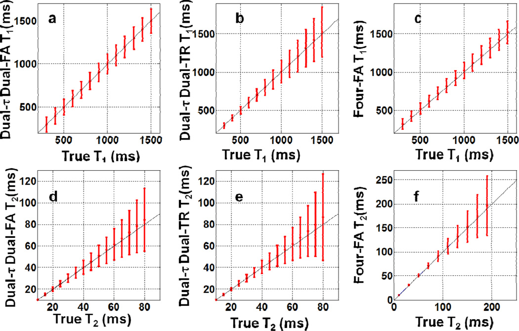
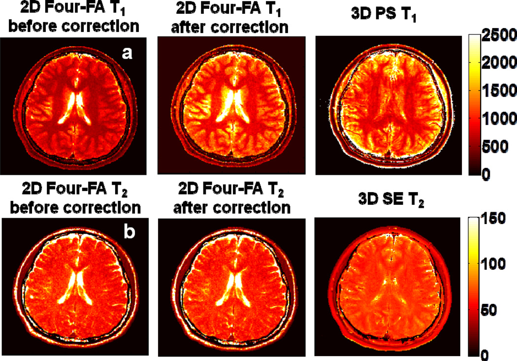
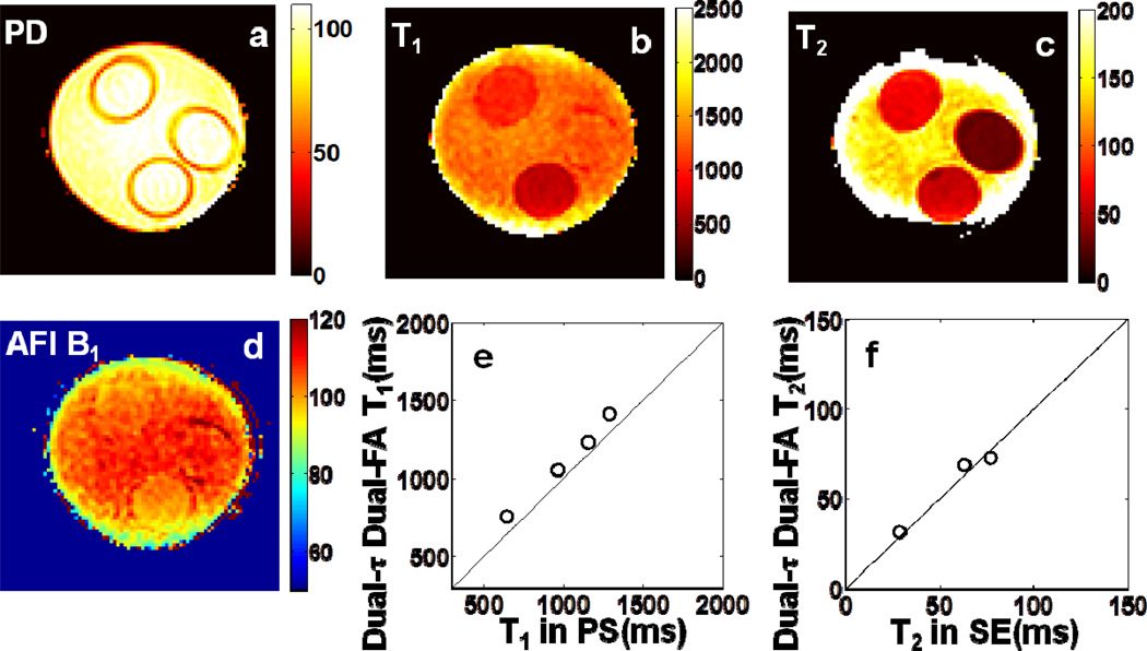
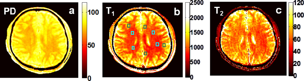
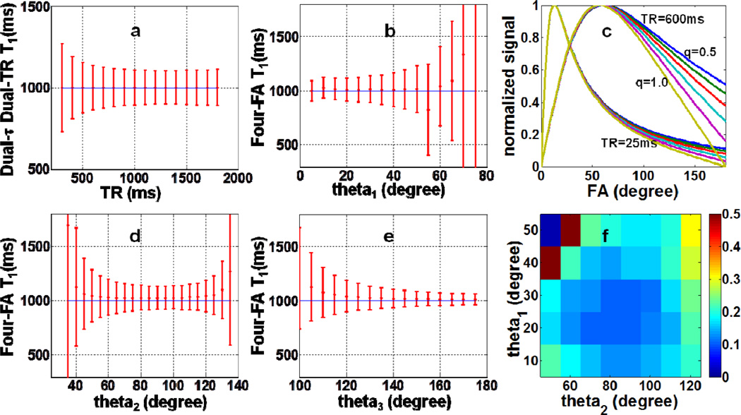
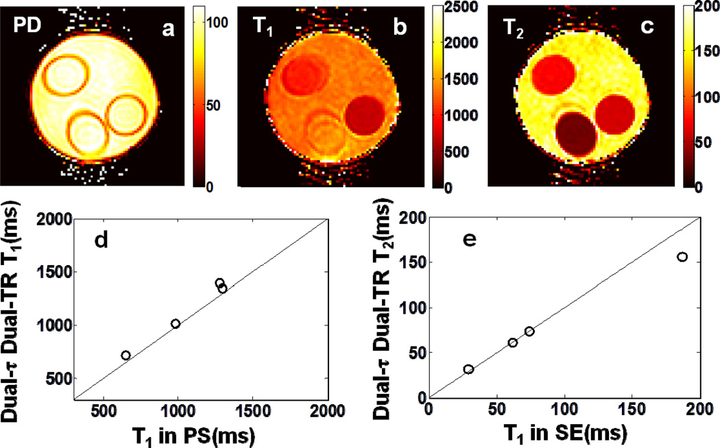
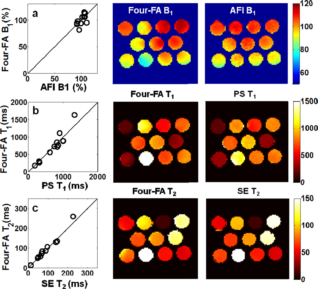
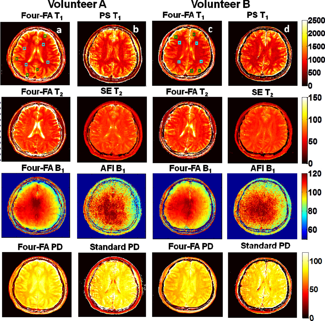
The spin lattice (T(1)) and spin-spin (T(2)) relaxation times, along with the proton density (PD) contain almost all of the information that (1)H MRI routinely uses in clinical diagnosis and research, but are seldom imaged directly. Here, three methods for directly imaging T(1), T(2), and PD with the least possible number of acquisitions - three, are presented. All methods utilize long 0° self-refocusing adiabatic pre-pulses instead of spin-echoes to encode the T(2) information prior to a conventional gradient-echo MRI sequence. T(1) information is encoded by varying the flip-angle (FA) in the 'Dual-τ Dual-FA' and 'Four-FA' methods, or the sequence repetition period, TR, in the 'Dual-τ Dual-TR' method. Inhomogeneity in the FA distribution and slice-selection profile are recognized as the main error sources for T(1) measurements. The former is remedied by integrating an extra FA-dependent acquisition into the 'Four-FA' method to provide self-corrected T(1), T(2), PD, and FA in just four acquisitions - again, the minimum possible. Slice profile errors - which manifest as differences between 2D and 3D T(1) measurements, can be addressed by Bloch equation analysis and experimental calibration. All three methods are validated in phantom studies, and the 'Dual-τ Dual-FA' and 'Four-FA' methods are validated in human brain studies using standard partial saturation and spin-echo methods for reference. The new methods offer a minimum-acquisition option for imaging single-component T(1), T(2), and PD. 'Four-FA' performs best overall in accuracy, with high efficiency per unit accuracy vs. existing methods when B(1)-inhomogeneity is appropriately addressed.
自旋晶格(T(1))和自旋 - 自旋(T(2))弛豫时间,以及质子密度(PD)几乎包含了氢质子磁共振成像(¹H MRI)在临床诊断和研究中常规使用的所有信息,但很少直接成像。本文介绍了三种以最少采集次数(三次)直接成像T(1)、T(2)和PD的方法。所有方法在传统梯度回波MRI序列之前,利用长0°自聚焦绝热预脉冲而非自旋回波来编码T(2)信息。在“双τ双翻转角(Dual-τ Dual-FA)”和“四翻转角(Four-FA)”方法中,通过改变翻转角(FA)来编码T(1)信息;在“双τ双重复时间(Dual-τ Dual-TR)”方法中,则通过改变序列重复时间TR来编码T(1)信息。翻转角分布的不均匀性和层面选择轮廓被认为是T(1)测量的主要误差来源。对于前者,通过在“四翻转角”方法中集成一个额外的与翻转角相关的采集来进行补救,从而仅通过四次采集(同样是最少可能次数)就能提供自校正的T(1)、T(2)、PD和FA。层面轮廓误差(表现为二维和三维T(1)测量之间的差异)可通过布洛赫方程分析和实验校准来解决。所有三种方法均在体模研究中得到验证,“双τ双翻转角”和“四翻转角”方法在人脑研究中也通过使用标准部分饱和和自旋回波方法作为参考得到了验证。这些新方法为单组分T(1)、T(2)和PD成像提供了最少采集次数的选择。当适当解决B(1)不均匀性问题时,“四翻转角”方法在准确性方面总体表现最佳,与现有方法相比,单位准确性下效率更高。