Harnish Roy, Prevrhal Sven, Alavi Abass, Zaidi Habib, Lang Thomas F
Department of Radiology and Biomedical Imaging, University of California, San Francisco, Box 0946, San Francisco, CA, 94143-0946, USA,
Ann Nucl Med. 2014 Jul;28(6):540-50. doi: 10.1007/s12149-014-0844-7. Epub 2014 Apr 8.
To determine if metal artefact reduction (MAR) combined with a priori knowledge of prosthesis material composition can be applied to obtain CT-based attenuation maps with sufficient accuracy for quantitative assessment of (18)F-fluorodeoxyglucose uptake in lesions near metallic prostheses.
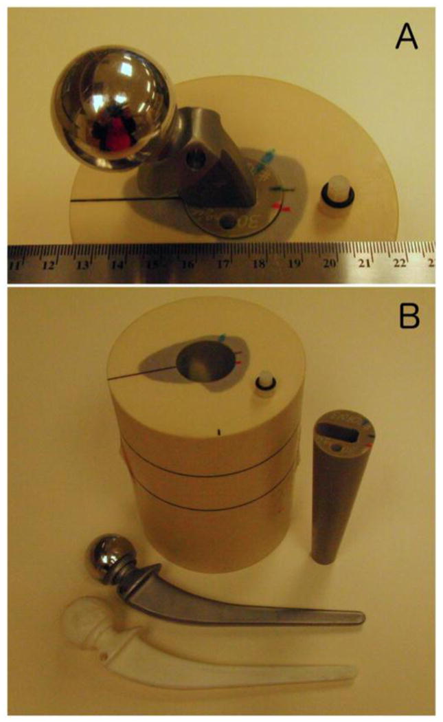
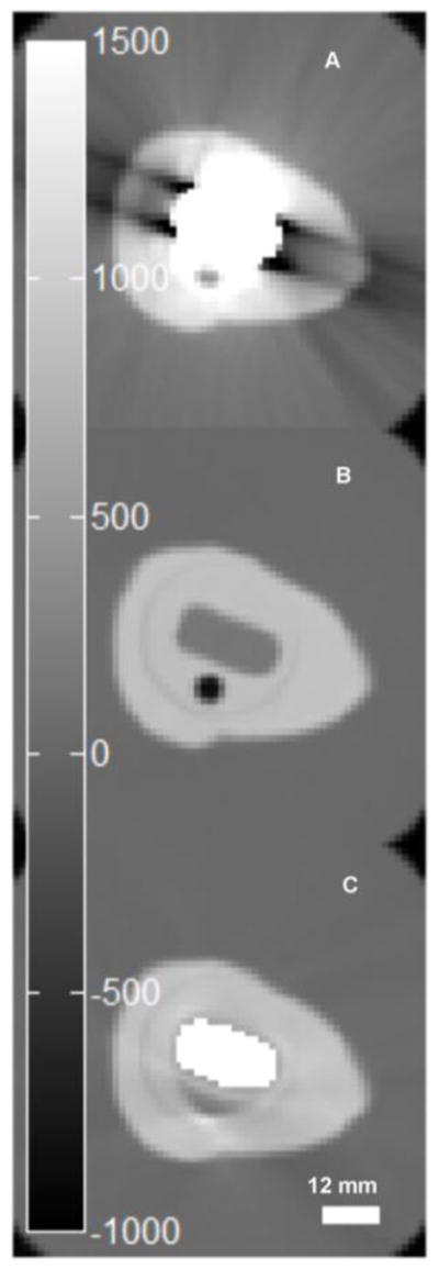
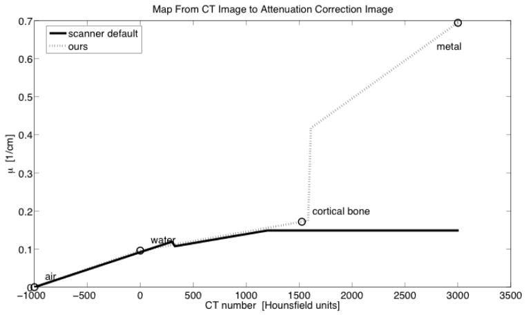
A custom hip prosthesis phantom with a lesion-sized cavity filled with 0.2 ml (18)F-FDG solution having an activity of 3.367 MBq adjacent to a prosthesis bore was imaged twice with a chrome-cobalt steel hip prosthesis and a plastic replica, respectively. Scanning was performed on a clinical hybrid PET/CT system equipped with an additional external (137)Cs transmission source. PET emission images were reconstructed from both phantom configurations with CT-based attenuation correction (CTAC) and with CT-based attenuation correction using MAR (MARCTAC). To compare results with the attenuation-correction method extant prior to the advent of PET/CT, we also carried out attenuation correction with (137)Cs transmission-based attenuation correction (TXAC). CTAC and MARCTAC images were scaled to attenuation coefficients at 511 keV using a trilinear function that mapped the highest CT values to the prosthesis alloy attenuation coefficient. Accuracy and spatial distribution of the lesion activity was compared between the three reconstruction schemes.
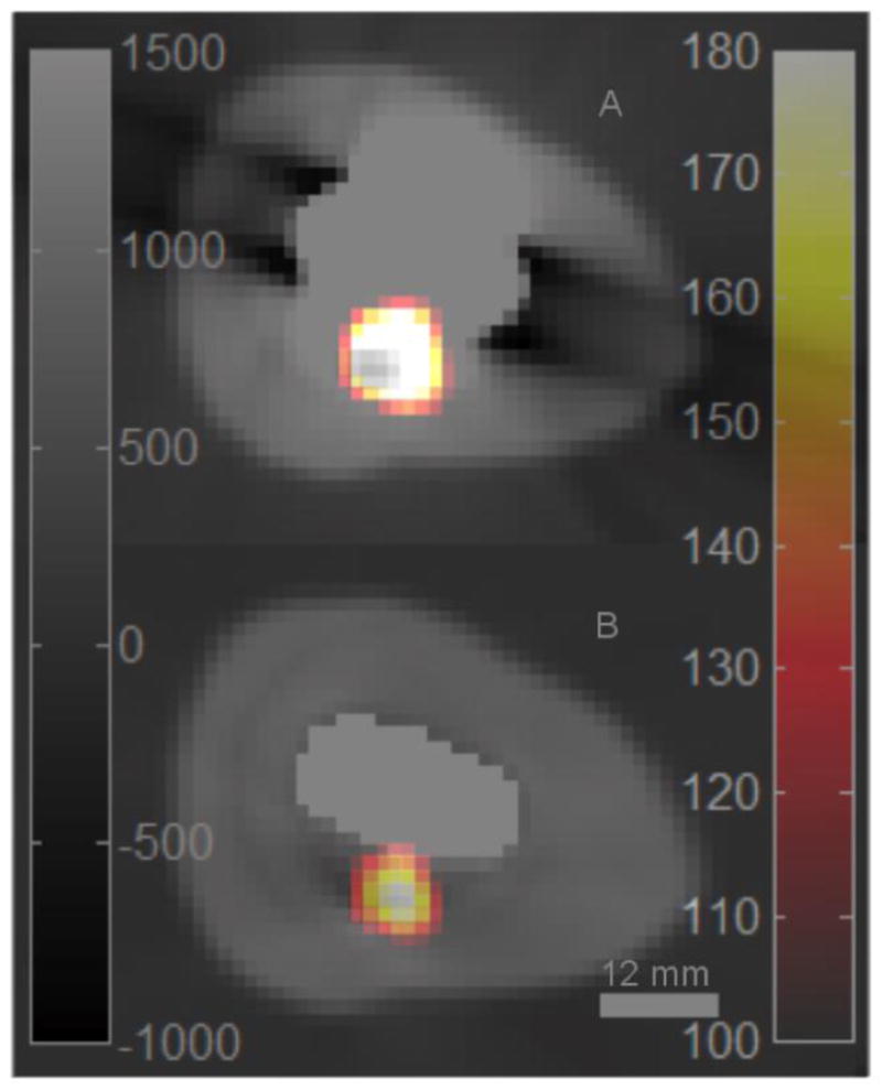
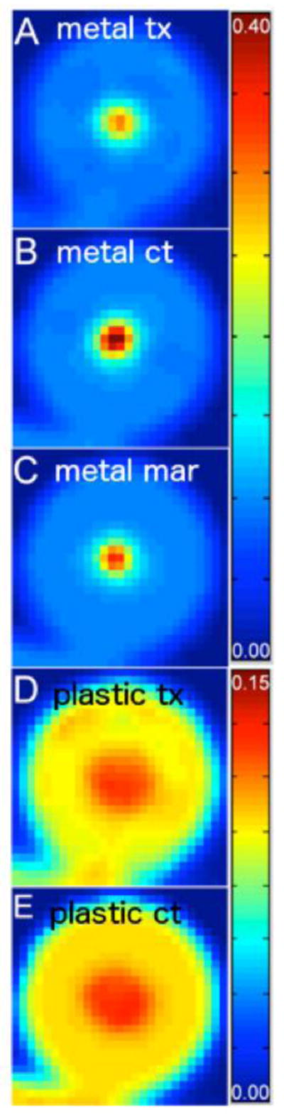
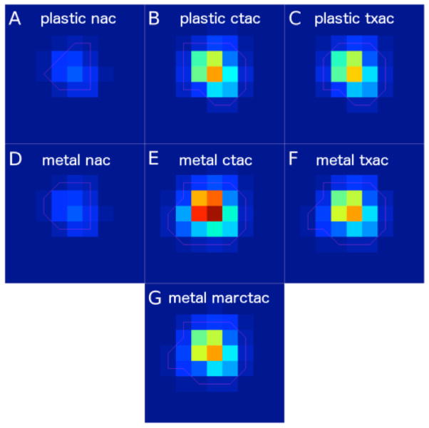
Compared to the reference activity of 3.37 MBq, the estimated activity quantified from the PET image corrected by TXAC was 3.41 MBq. The activity estimated from PET images corrected by MARCTAC was similar in accuracy at 3.32 MBq. CTAC corrected PET images resulted in nearly 40 % overestimation of lesion activity at 4.70 MBq. Comparison of PET images obtained with the plastic and metal prostheses in place showed that CTAC resulted in a marked distortion of the (18)F-FDG distribution within the lesion, whereas application of MARCTAC and TXAC resulted in lesion distributions similar to those observed with the plastic replica.
MAR combined with a trilinear CT number mapping for PET attenuation correction resulted in estimates of lesion activity comparable in accuracy to that obtained with (137)Cs transmission-based attenuation correction, and far superior to estimates made without attenuation correction or with a standard CT attenuation map. The ability to use CT images for attenuation correction is a potentially important development because it obviates the need for a (137)Cs transmission source, which entails extra scan time, logistical complexity and expense.
确定金属伪影减少(MAR)与假体材料成分的先验知识相结合是否可用于获得基于CT的衰减图,其精度足以对金属假体附近病变中的(18)F-氟脱氧葡萄糖摄取进行定量评估。
一个定制的髋关节假体模型,在假体孔附近有一个病变大小的腔,腔内填充0.2 ml活度为3.367 MBq的(18)F-FDG溶液,分别用铬钴钢髋关节假体和塑料复制品对其进行了两次成像。在配备额外外部(137)Cs透射源的临床混合型PET/CT系统上进行扫描。PET发射图像通过基于CT的衰减校正(CTAC)以及使用MAR的基于CT的衰减校正(MARCTAC)从两种模型配置重建。为了将结果与PET/CT出现之前存在的衰减校正方法进行比较,我们还使用基于(137)Cs透射的衰减校正(TXAC)进行了衰减校正。使用将最高CT值映射到假体合金衰减系数的三线函数,将CTAC和MARCTAC图像缩放到511 keV处的衰减系数。比较三种重建方案之间病变活性的准确性和空间分布。
与参考活度3.37 MBq相比,通过TXAC校正的PET图像定量估计的活度为3.41 MBq。通过MARCTAC校正的PET图像估计的活度在3.32 MBq时准确性相似。CTAC校正的PET图像导致病变活性高估近40%,为4.70 MBq。对放置塑料和金属假体时获得的PET图像进行比较表明,CTAC导致病变内(18)F-FDG分布明显扭曲,而应用MARCTAC和TXAC导致病变分布与塑料复制品观察到的相似。
MAR与用于PET衰减校正的三线CT值映射相结合,导致病变活性估计的准确性与基于(137)Cs透射的衰减校正相当,并且远优于未进行衰减校正或使用标准CT衰减图的估计。使用CT图像进行衰减校正的能力是一个潜在的重要进展,因为它无需(137)Cs透射源,而该源需要额外的扫描时间、后勤复杂性和费用。