Division of Nuclear Medicine and Molecular Imaging, Geneva University Hospital, CH-1211, Geneva 4, Switzerland.
Geneva University Neurocenter, Geneva University, CH-1205, Geneva, Switzerland.
Eur Radiol. 2021 Aug;31(8):6384-6396. doi: 10.1007/s00330-021-07709-z. Epub 2021 Feb 10.
The susceptibility of CT imaging to metallic objects gives rise to strong streak artefacts and skewed information about the attenuation medium around the metallic implants. This metal-induced artefact in CT images leads to inaccurate attenuation correction in PET/CT imaging. This study investigates the potential of deep learning-based metal artefact reduction (MAR) in quantitative PET/CT imaging.
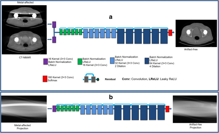
Deep learning-based metal artefact reduction approaches were implemented in the image (DLI-MAR) and projection (DLP-MAR) domains. The proposed algorithms were quantitatively compared to the normalized MAR (NMAR) method using simulated and clinical studies. Eighty metal-free CT images were employed for simulation of metal artefact as well as training and evaluation of the aforementioned MAR approaches. Thirty F-FDG PET/CT images affected by the presence of metallic implants were retrospectively employed for clinical assessment of the MAR techniques.
The evaluation of MAR techniques on the simulation dataset demonstrated the superior performance of the DLI-MAR approach (structural similarity (SSIM) = 0.95 ± 0.2 compared to 0.94 ± 0.2 and 0.93 ± 0.3 obtained using DLP-MAR and NMAR, respectively) in minimizing metal artefacts in CT images. The presence of metallic artefacts in CT images or PET attenuation correction maps led to quantitative bias, image artefacts and under- and overestimation of scatter correction of PET images. The DLI-MAR technique led to a quantitative PET bias of 1.3 ± 3% compared to 10.5 ± 6% without MAR and 3.2 ± 0.5% achieved by NMAR.
The DLI-MAR technique was able to reduce the adverse effects of metal artefacts on PET images through the generation of accurate attenuation maps from corrupted CT images.
• The presence of metallic objects, such as dental implants, gives rise to severe photon starvation, beam hardening and scattering, thus leading to adverse artefacts in reconstructed CT images. • The aim of this work is to develop and evaluate a deep learning-based MAR to improve CT-based attenuation and scatter correction in PET/CT imaging. • Deep learning-based MAR in the image (DLI-MAR) domain outperformed its counterpart implemented in the projection (DLP-MAR) domain. The DLI-MAR approach minimized the adverse impact of metal artefacts on whole-body PET images through generating accurate attenuation maps from corrupted CT images.
CT 成像对金属物体的敏感性会产生强烈的条纹伪影,并对金属植入物周围的衰减介质产生倾斜的信息。CT 图像中的这种金属诱导的伪影会导致 PET/CT 成像中的衰减校正不准确。本研究探讨了基于深度学习的金属伪影减少(MAR)在定量 PET/CT 成像中的潜力。
在图像(DLI-MAR)和投影(DLP-MAR)域中实现了基于深度学习的金属伪影减少方法。使用模拟和临床研究,将所提出的算法与归一化 MAR(NMAR)方法进行了定量比较。使用 80 张无金属 CT 图像模拟金属伪影,并对上述 MAR 方法进行培训和评估。回顾性地使用 30 张受金属植入物影响的 F-FDG PET/CT 图像来评估 MAR 技术的临床效果。
在模拟数据集上评估 MAR 技术的结果表明,DLI-MAR 方法的性能优于 DLP-MAR 和 NMAR(结构相似性(SSIM)分别为 0.95±0.2、0.94±0.2 和 0.93±0.3),可最大程度地减少 CT 图像中的金属伪影。CT 图像或 PET 衰减校正图中的金属伪影会导致定量偏差、图像伪影以及 PET 散射校正的低估和高估。与未经 MAR 和 NMAR 处理的 10.5±6%和 3.2±0.5%相比,DLI-MAR 技术导致 PET 定量偏差为 1.3±3%。
通过从受损 CT 图像生成准确的衰减图,DLI-MAR 技术能够减少金属伪影对 PET 图像的不利影响。
• 金属物体(如牙植入物)的存在会导致严重的光子饥饿、束硬化和散射,从而导致重建 CT 图像中的不良伪影。• 本工作的目的是开发和评估一种基于深度学习的 MAR,以改善 PET/CT 成像中的 CT 基衰减和散射校正。• 在图像(DLI-MAR)域中实现的基于深度学习的 MAR 优于在投影(DLP-MAR)域中实现的 MAR。DLI-MAR 方法通过从受损 CT 图像生成准确的衰减图,最大程度地减少了金属伪影对全身 PET 图像的不利影响。