Robinson Scott M, Tsueng Ginger, Sin Jon, Mangale Vrushali, Rahawi Shahad, McIntyre Laura L, Williams Wesley, Kha Nelson, Cruz Casey, Hancock Bryan M, Nguyen David P, Sayen M Richard, Hilton Brett J, Doran Kelly S, Segall Anca M, Wolkowicz Roland, Cornell Christopher T, Whitton J Lindsay, Gottlieb Roberta A, Feuer Ralph
The Integrated Regenerative Research Institute (IRRI) at San Diego State University, Cell & Molecular Biology Joint Doctoral Program, Department of Biology, San Diego State University, San Diego, California, United States of America.
Donald P. Shiley BioScience Center, San Diego State University, San Diego, California, United States of America.
PLoS Pathog. 2014 Apr 10;10(4):e1004045. doi: 10.1371/journal.ppat.1004045. eCollection 2014 Apr.
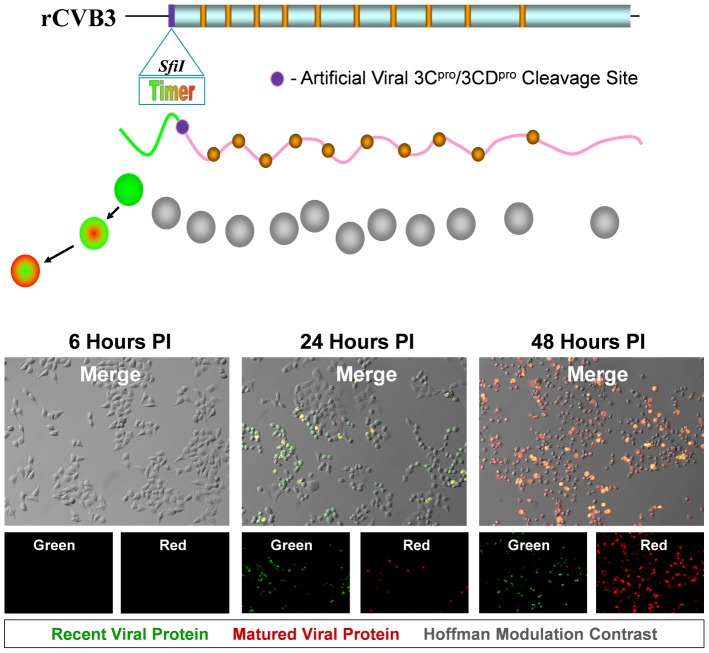
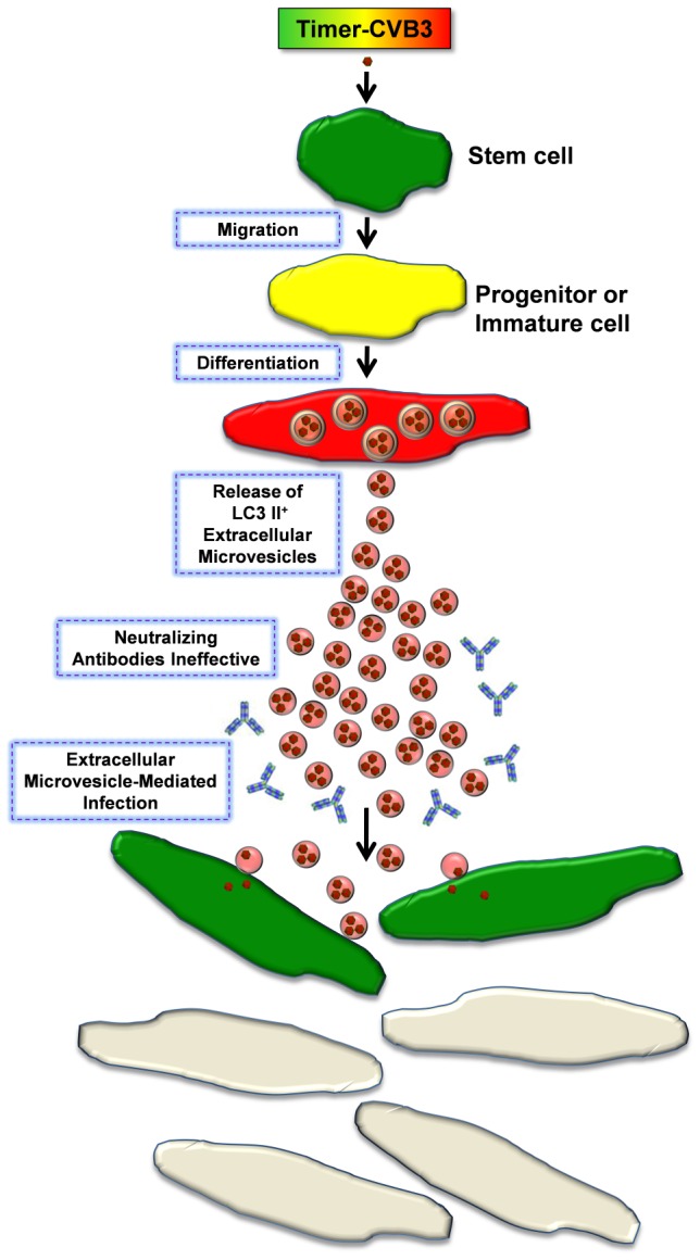
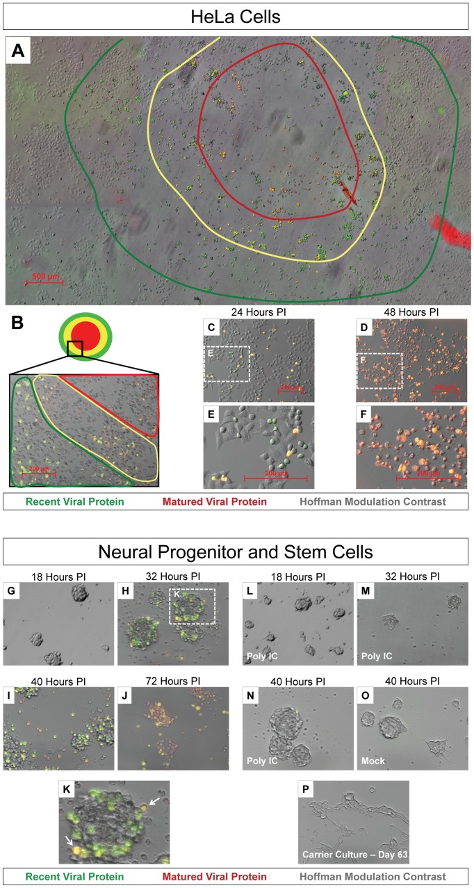
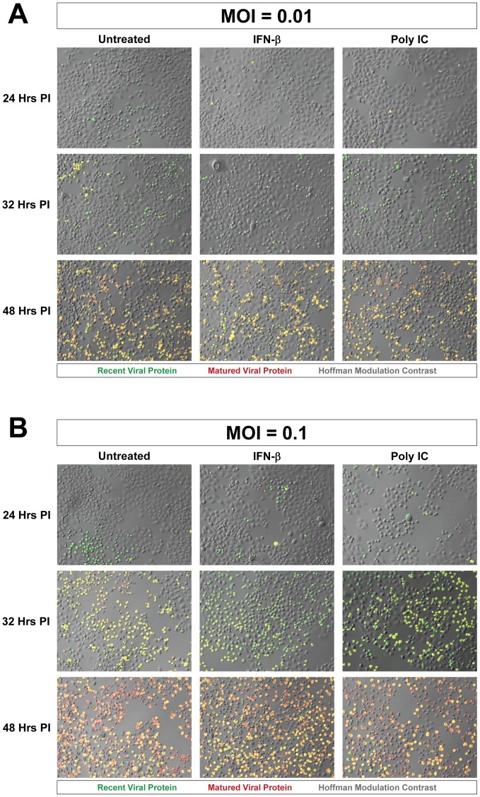
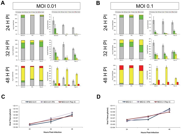
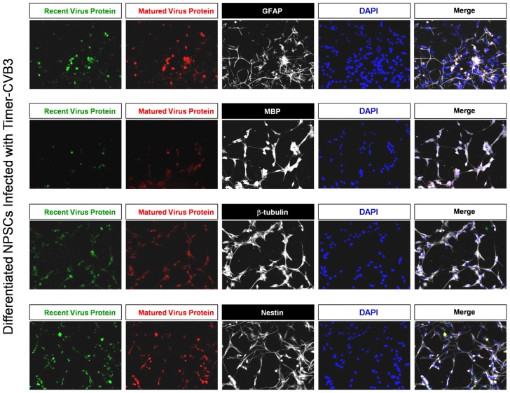
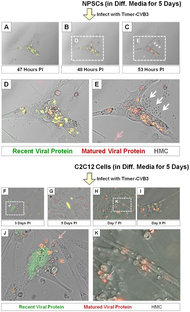
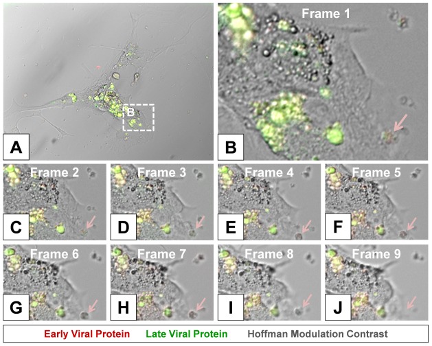
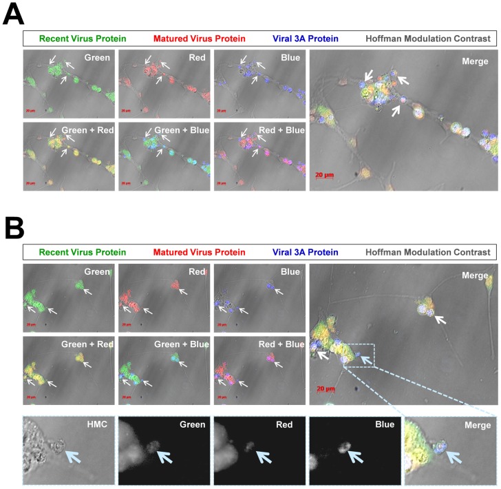
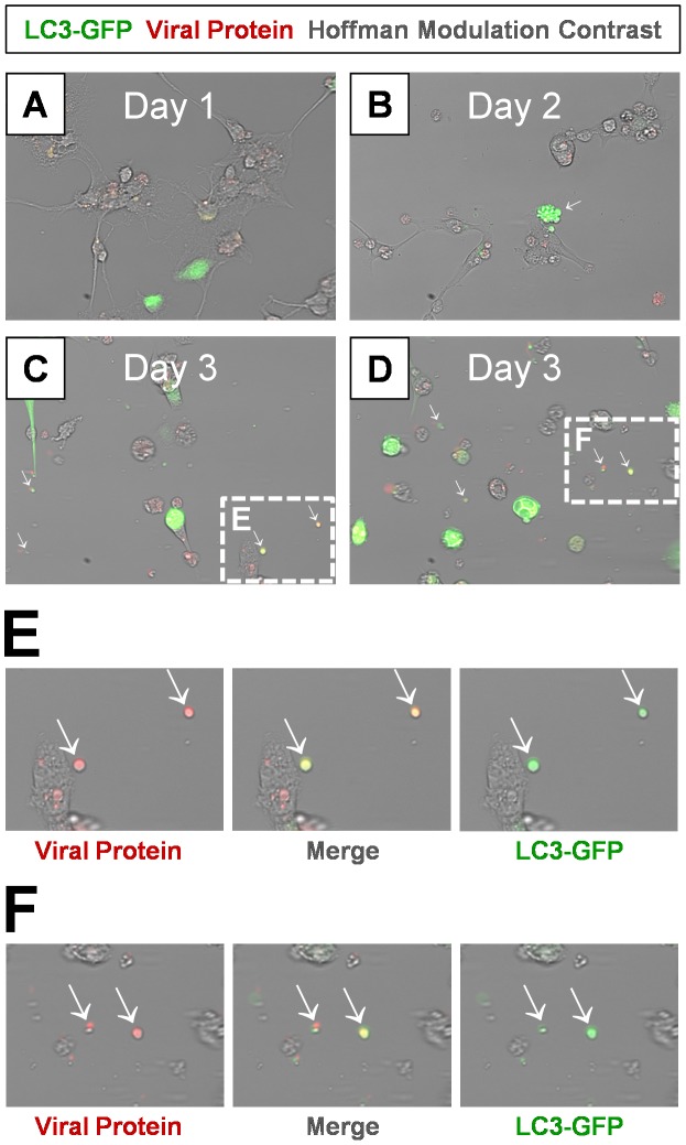
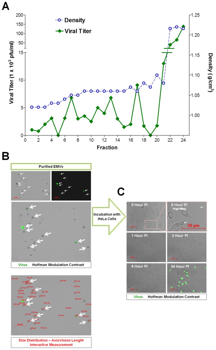
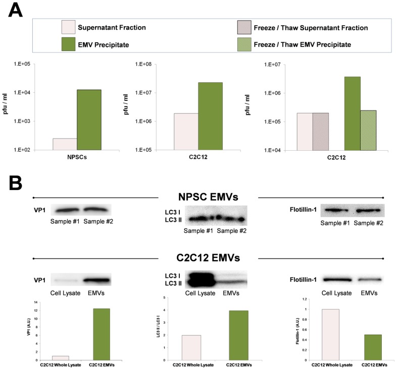
Coxsackievirus B3 (CVB3), a member of the picornavirus family and enterovirus genus, causes viral myocarditis, aseptic meningitis, and pancreatitis in humans. We genetically engineered a unique molecular marker, "fluorescent timer" protein, within our infectious CVB3 clone and isolated a high-titer recombinant viral stock (Timer-CVB3) following transfection in HeLa cells. "Fluorescent timer" protein undergoes slow conversion of fluorescence from green to red over time, and Timer-CVB3 can be utilized to track virus infection and dissemination in real time. Upon infection with Timer-CVB3, HeLa cells, neural progenitor and stem cells (NPSCs), and C2C12 myoblast cells slowly changed fluorescence from green to red over 72 hours as determined by fluorescence microscopy or flow cytometric analysis. The conversion of "fluorescent timer" protein in HeLa cells infected with Timer-CVB3 could be interrupted by fixation, suggesting that the fluorophore was stabilized by formaldehyde cross-linking reactions. Induction of a type I interferon response or ribavirin treatment reduced the progression of cell-to-cell virus spread in HeLa cells or NPSCs infected with Timer-CVB3. Time lapse photography of partially differentiated NPSCs infected with Timer-CVB3 revealed substantial intracellular membrane remodeling and the assembly of discrete virus replication organelles which changed fluorescence color in an asynchronous fashion within the cell. "Fluorescent timer" protein colocalized closely with viral 3A protein within virus replication organelles. Intriguingly, infection of partially differentiated NPSCs or C2C12 myoblast cells induced the release of abundant extracellular microvesicles (EMVs) containing matured "fluorescent timer" protein and infectious virus representing a novel route of virus dissemination. CVB3 virions were readily observed within purified EMVs by transmission electron microscopy, and infectious virus was identified within low-density isopycnic iodixanol gradient fractions consistent with membrane association. The preferential detection of the lipidated form of LC3 protein (LC3 II) in released EMVs harboring infectious virus suggests that the autophagy pathway plays a crucial role in microvesicle shedding and virus release, similar to a process previously described as autophagosome-mediated exit without lysis (AWOL) observed during poliovirus replication. Through the use of this novel recombinant virus which provides more dynamic information from static fluorescent images, we hope to gain a better understanding of CVB3 tropism, intracellular membrane reorganization, and virus-associated microvesicle dissemination within the host.
柯萨奇病毒B3(CVB3)是小RNA病毒科肠道病毒属的成员,可导致人类病毒性心肌炎、无菌性脑膜炎和胰腺炎。我们在具有感染性的CVB3克隆中通过基因工程改造了一种独特的分子标记——“荧光定时器”蛋白,并在HeLa细胞中转染后分离出了高滴度的重组病毒株(Timer-CVB3)。“荧光定时器”蛋白的荧光会随着时间缓慢地从绿色转变为红色,Timer-CVB3可用于实时追踪病毒的感染和传播。用Timer-CVB3感染后,通过荧光显微镜或流式细胞术分析可知,HeLa细胞、神经祖细胞和干细胞(NPSCs)以及C2C12成肌细胞在72小时内荧光从绿色缓慢转变为红色。感染Timer-CVB3的HeLa细胞中“荧光定时器”蛋白的转变可被固定所阻断,这表明荧光团通过甲醛交联反应得以稳定。I型干扰素应答的诱导或利巴韦林处理可减少感染Timer-CVB3的HeLa细胞或NPSCs中细胞间病毒传播的进程。对感染Timer-CVB3的部分分化NPSCs进行延时摄影,发现细胞内有大量的内膜重塑以及离散的病毒复制细胞器的组装,这些细胞器在细胞内以异步方式改变荧光颜色。“荧光定时器”蛋白与病毒复制细胞器内的病毒3A蛋白紧密共定位。有趣的是,感染部分分化的NPSCs或C2C12成肌细胞会诱导释放大量含有成熟“荧光定时器”蛋白和感染性病毒的细胞外微泡(EMV),这代表了一种新的病毒传播途径。通过透射电子显微镜在纯化的EMV中很容易观察到CVB3病毒粒子,并且在与膜相关的低密度等密度碘克沙醇梯度组分中鉴定出了感染性病毒。在含有感染性病毒的释放的EMV中优先检测到脂化形式的LC3蛋白(LC3 II),这表明自噬途径在微泡脱落和病毒释放中起关键作用,类似于脊髓灰质炎病毒复制过程中先前描述的自噬体介导的不裂解退出(AWOL)过程。通过使用这种新型重组病毒,它能从静态荧光图像中提供更多动态信息,我们希望能更好地了解CVB3的嗜性、细胞内膜重组以及病毒相关微泡在宿主体内的传播。