Luna-Cancalon Katiuska, Sikora Kristine M, Pappas Samuel S, Singh Vikrant, Wulff Heike, Paulson Henry L, Burmeister Margit, Shakkottai Vikram G
Department of Neurology, University of Michigan, Ann Arbor, MI 48109, USA.
Program in Cellular and Molecular Biology, University of Michigan, Ann Arbor, MI 48109, USA.
Neurobiol Dis. 2014 Jul;67:140-8. doi: 10.1016/j.nbd.2014.03.020. Epub 2014 Apr 12.
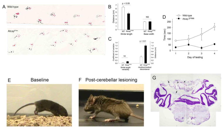
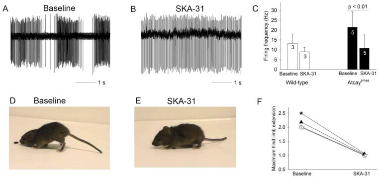
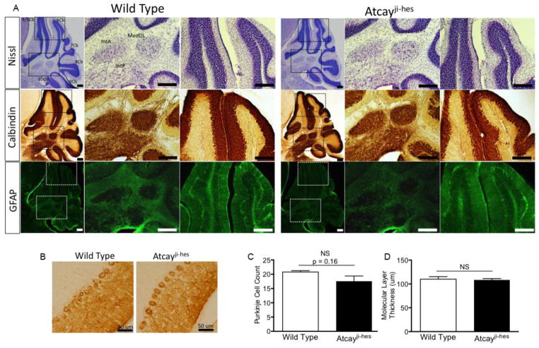
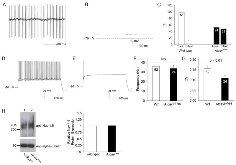
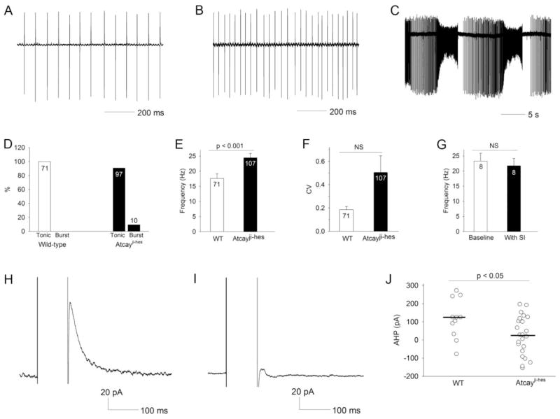
Recent evidence suggests that dystonia, a movement disorder characterized by sustained involuntary muscle contractions, can be associated with cerebellar abnormalities. The basis for how functional changes in the cerebellum can cause dystonia is poorly understood. Here we identify alterations in physiology in Atcay(ji-hes) mice which in addition to ataxia, have an abnormal gait with hind limb extension and toe walking, reminiscent of human dystonic gait. No morphological abnormalities in the brain accompany the dystonia, but partial cerebellectomy causes resolution of the stiff-legged gait, suggesting that cerebellar dysfunction contributes to the dystonic gait of Atcay(ji-hes) mice. Recordings from Purkinje and deep cerebellar nuclear (DCN) neurons in acute brain slices were used to determine the physiological correlates of dystonia in the Atcay(ji-hes) mice. Approximately 50% of cerebellar Purkinje neurons fail to display the normal repetitive firing characteristic of these cells. In addition, DCN neurons exhibit increased intrinsic firing frequencies with a subset of neurons displaying bursts of action potentials. This increased intrinsic excitability of DCN neurons is accompanied by a reduction in after-hyperpolarization currents mediated by small-conductance calcium-activated potassium (SK) channels. An activator of SK channels reduces DCN neuron firing frequency in acute cerebellar slices and improves the dystonic gait of Atcay(ji-hes) mice. These results suggest that a combination of reduced Purkinje neuron activity and increased DCN intrinsic excitability can result in a combination of ataxia and a dystonia-like gait in mice.
近期证据表明,肌张力障碍作为一种以持续性非自愿肌肉收缩为特征的运动障碍,可能与小脑异常有关。小脑的功能变化如何导致肌张力障碍,目前尚不清楚。在这里,我们发现了Atcay(ji - hes)小鼠的生理变化,这些小鼠除了共济失调外,还具有异常步态,表现为后肢伸展和足尖行走,类似于人类的肌张力障碍步态。肌张力障碍出现时,大脑没有形态学异常,但部分小脑切除可使僵硬的腿部步态得到缓解,这表明小脑功能障碍导致了Atcay(ji - hes)小鼠的肌张力障碍步态。通过对急性脑片中小脑浦肯野神经元和小脑深部核团(DCN)神经元的记录,来确定Atcay(ji - hes)小鼠肌张力障碍的生理相关性。大约50%的小脑浦肯野神经元未能表现出这些细胞正常的重复放电特征。此外,DCN神经元的内在放电频率增加,一部分神经元表现出动作电位爆发。DCN神经元内在兴奋性的增加伴随着小电导钙激活钾(SK)通道介导的超极化后电流的减少。SK通道激活剂可降低急性小脑切片中DCN神经元的放电频率,并改善Atcay(ji - hes)小鼠的肌张力障碍步态。这些结果表明,浦肯野神经元活动减少和DCN内在兴奋性增加共同作用,可导致小鼠出现共济失调和类似肌张力障碍的步态。