Division of Breast and Endocrine Surgery, Department of Surgery, Samsung Medical Center, Sungkyunkwan University School of Medicine, Seoul, Korea.
Department of Surgery, Soonchunhyang University Cheonan Hospital, Soonchunhyang University College of Medicine, Cheonan, Korea.
J Breast Cancer. 2014 Mar;17(1):83-7. doi: 10.4048/jbc.2014.17.1.83. Epub 2014 Mar 28.
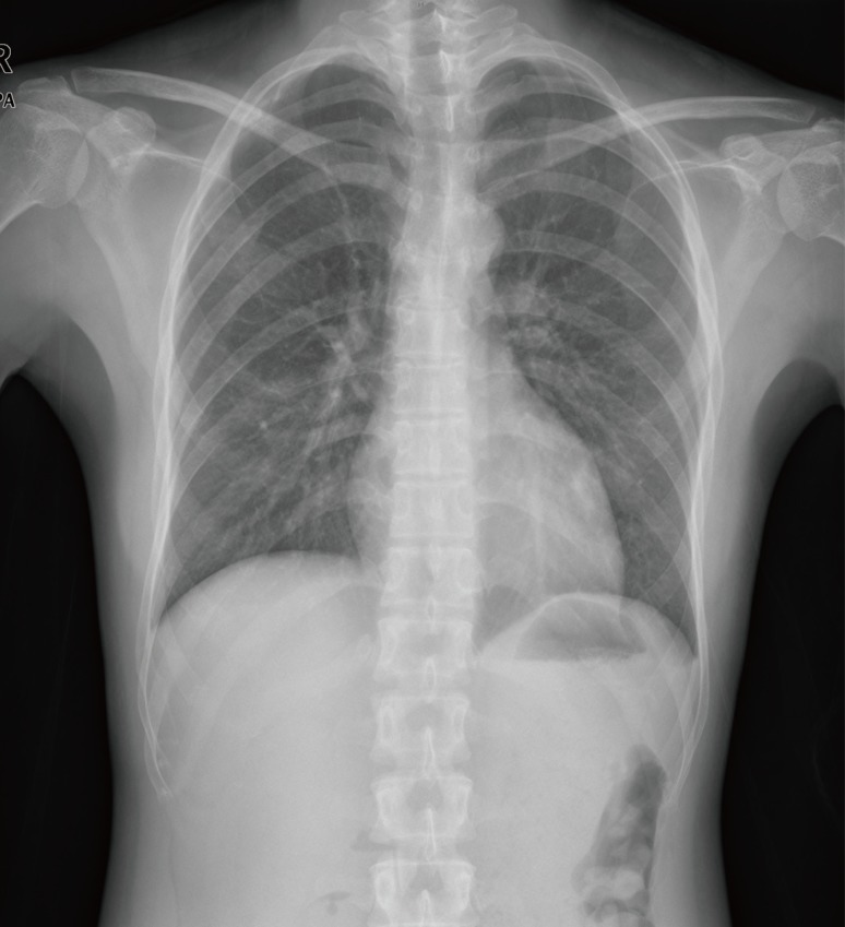
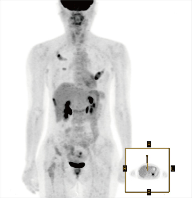
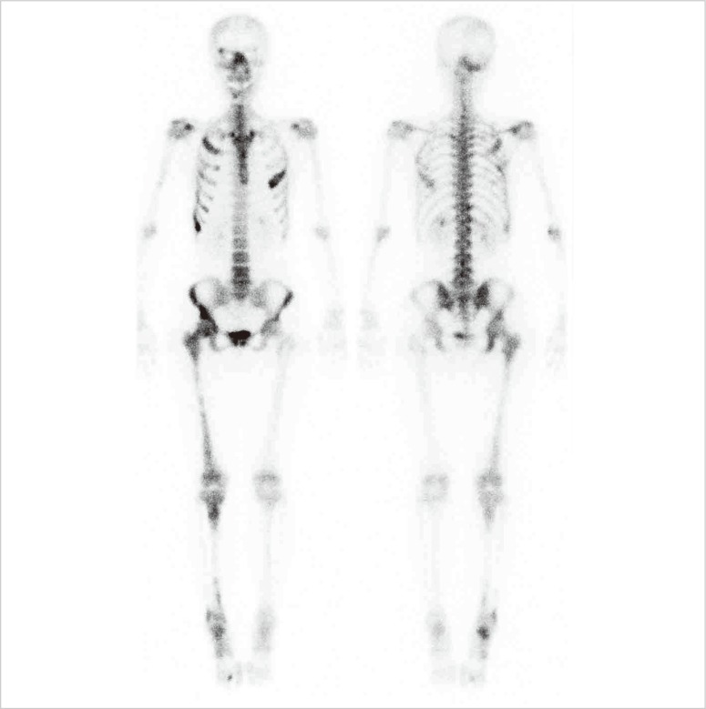
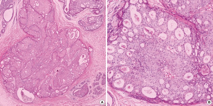
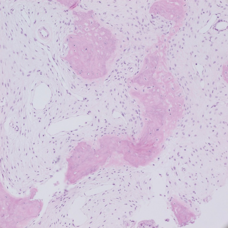
Whole-body bone scans and whole body (18)F-fluorodeoxyglucose positron emission tomographic/computed tomographic scans are sensitive for detecting bone metastasis in patients with breast cancer. However, it is often difficult to discriminate between bone metastasis and other nonmalignant bone lesions. Polyostotic fibrous dysplasia is a rare disorder characterized by the osteoid medullary cavity filling with fibrous tissue causing bony expansion. We report the case of a 42-year-old female patient with ductal carcinoma in situ, which appeared to have multiple bone metastases on initial work-up images. Subsequently, the bone metastases were identified as polyostotic fibrous dysplasia. The patient underwent modified radical mastectomy and subsequently visited for a second opinion regarding the bony metastases. She underwent right ilium computed tomography-guided biopsy. Pathology was consistent with fibrous dysplasia. This patient received only adjuvant tamoxifen, and 1.5 years later, there was no evidence of recurrence.
全身骨扫描和全身(18)F-氟代脱氧葡萄糖正电子发射断层扫描/计算机断层扫描对乳腺癌患者的骨转移具有较高的敏感性。然而,通常很难区分骨转移和其他非恶性骨病变。多灶性纤维结构不良是一种罕见的疾病,其特征是骨样骨髓腔充满纤维组织导致骨膨胀。我们报告了一例 42 岁女性患者,患有原位导管癌,初步检查图像显示多处骨转移。随后,骨转移被确定为多灶性纤维结构不良。患者接受了改良根治性乳房切除术,随后因骨转移而来进行第二次咨询。她接受了右侧髂骨 CT 引导下活检。病理与纤维结构不良一致。该患者仅接受了辅助他莫昔芬治疗,1.5 年后,没有复发的证据。