Yamaue Yasuhiro, Hosaka Yoshinao Z, Uehara Masato
Department of Veterinary Anatomy, Faculty of Agriculture, Tottori University, Tottori 680-8553, Japan.
J Vet Med Sci. 2014 Aug;76(8):1099-103. doi: 10.1292/jvms.14-0132. Epub 2014 Apr 30.
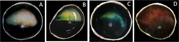
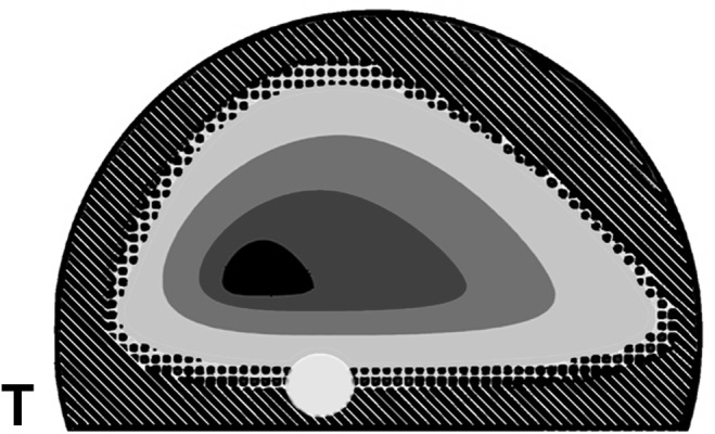
We aimed to document macroscopic variations in the cellular tapetum in the dog, to provide a histologic description of the macroscopic results and to evaluate the correlation between the macroscopic appearance and aging. Fifty three dogs including 5 beagles, 1 Chihuahua and 47 mixed breeds of each gender were used. For a macroscopic study, the fresh tapetal fundi were photographed using digital camera. For a histological study, the glutaraldehyde-formalin fixed eyes were embedded in nitrocellulose and stained with hematoxylin-eosin or thionine. The normal tapetum was triangular with the rounded angles and the smooth contour. The atypical tapetum was smaller and more variable in shape, contour and color than the normal one. In severe cases, the fundus was devoid of the tapetum. The atypical tapetum tended to increase in frequency with aging. Retinal pigment epithelial cells on the normal tapetum were unpigmented. In the eye with the atypical tapetum, regardless of tapetal size and shape, unpigmented retinal pigment epithelial cells showed a similar distribution to that on the normal tapetum, even in a dog without a tapetum. Although there is a congenitally hypoplastic tapetum, the atypical tapetum tends to increase in incidence and severity with aging.
我们旨在记录犬类细胞性脉络膜毯的宏观变化,对宏观结果进行组织学描述,并评估宏观外观与衰老之间的相关性。使用了53只犬,包括5只比格犬、1只吉娃娃犬和47只各性别的混种犬。对于宏观研究,使用数码相机拍摄新鲜的脉络膜毯眼底。对于组织学研究,将戊二醛 - 福尔马林固定的眼睛包埋在硝化纤维素中,并用苏木精 - 伊红或硫堇染色。正常的脉络膜毯呈三角形,角圆钝,轮廓光滑。非典型脉络膜毯比正常的更小,形状、轮廓和颜色变化更大。严重情况下,眼底没有脉络膜毯。非典型脉络膜毯的出现频率往往随衰老而增加。正常脉络膜毯上的视网膜色素上皮细胞无色素沉着。在有非典型脉络膜毯的眼睛中,无论脉络膜毯的大小和形状如何,无色素沉着的视网膜色素上皮细胞的分布与正常脉络膜毯上的相似,即使在没有脉络膜毯的犬眼中也是如此。尽管存在先天性发育不全的脉络膜毯,但非典型脉络膜毯的发生率和严重程度往往随衰老而增加。