Akil Omar, Hall-Glenn Faith, Chang Jolie, Li Alfred, Chang Wenhan, Lustig Lawrence R, Alliston Tamara, Hsiao Edward C
Department of Otolaryngology, Head & Neck Surgery, University of California San Francisco, San Francisco, California, United States of America.
Department of Orthopaedic Surgery, University of California San Francisco, San Francisco, California, United States of America.
PLoS One. 2014 May 1;9(5):e94989. doi: 10.1371/journal.pone.0094989. eCollection 2014.
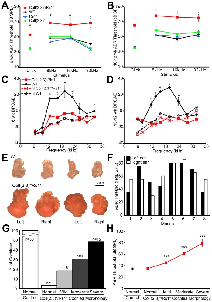
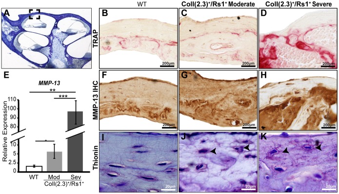
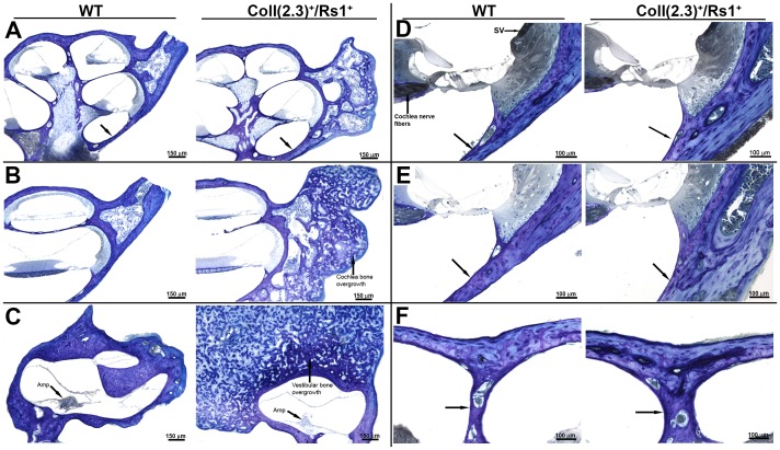
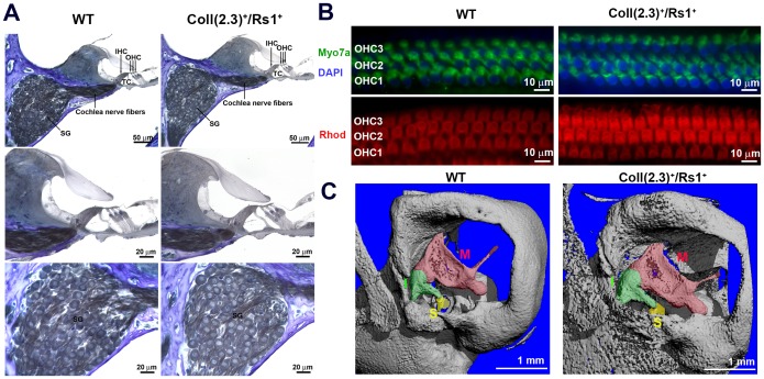
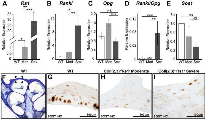
Normal hearing requires exquisite cooperation between bony and sensorineural structures within the cochlea. For example, the inner ear secretes proteins such as osteoprotegrin (OPG) that can prevent cochlear bone remodeling. Accordingly, diseases that affect bone regulation can also result in hearing loss. Patients with fibrous dysplasia develop trabecular bone overgrowth resulting in hearing loss if the lesions affect the temporal bones. Unfortunately, the mechanisms responsible for this hearing loss, which could be sensorineural and/or conductive, remain unclear. In this study, we used a unique transgenic mouse model of increased Gs G-protein coupled receptor (GPCR) signaling induced by expression of an engineered receptor, Rs1, in osteoblastic cells. These ColI(2.3)+/Rs1+ mice showed dramatic bone lesions that histologically and radiologically resembled fibrous dysplasia. We found that ColI(2.3)+/Rs1+ mice showed progressive and severe conductive hearing loss. Ossicular chain impingement increased with the size and number of dysplastic lesions. While sensorineural structures were unaffected, ColI(2.3)+/Rs1+ cochleae had abnormally high osteoclast activity, together with elevated tartrate resistant acid phosphatase (TRAP) activity and receptor activator of nuclear factor kappa-B ligand (Rankl) mRNA expression. ColI(2.3)+/Rs1+ cochleae also showed decreased expression of Sclerostin (Sost), an antagonist of the Wnt signaling pathway that normally increases bone formation. The osteocyte canalicular networks of ColI(2.3)+/Rs1+ cochleae were disrupted and showed abnormal osteocyte morphology. The osteocytes in the ColI(2.3)+/Rs1+ cochleae showed increased expression of matrix metalloproteinase 13 (MMP-13) and TRAP, both of which can support osteocyte-mediated peri-lacunar remodeling. Thus, while the ossicular chain impingement is sufficient to account for the progressive hearing loss in fibrous dysplasia, the deregulation of bone remodeling extends to the cochlea as well. Our findings suggest that factors regulating bone remodeling, including peri-lacunar remodeling by osteocytes, may be useful targets for treating the bony overgrowths and hearing changes of fibrous dysplasia and other bony pathologies.
正常听力需要耳蜗内骨结构和感觉神经结构之间的精确协作。例如,内耳会分泌骨保护素(OPG)等蛋白质,这些蛋白质可以防止耳蜗骨重塑。因此,影响骨调节的疾病也可能导致听力损失。患有纤维性发育不良的患者会出现小梁骨过度生长,如果病变影响颞骨,就会导致听力损失。不幸的是,这种听力损失的机制,可能是感觉神经性的和/或传导性的,仍然不清楚。在本研究中,我们使用了一种独特的转基因小鼠模型,该模型通过在成骨细胞中表达工程受体Rs1来增加Gs G蛋白偶联受体(GPCR)信号传导。这些ColI(2.3)+/Rs1+小鼠表现出明显的骨病变,在组织学和放射学上类似于纤维性发育不良。我们发现ColI(2.3)+/Rs1+小鼠表现出进行性和严重的传导性听力损失。听骨链受压随着发育异常病变的大小和数量增加而加重。虽然感觉神经结构未受影响,但ColI(2.3)+/Rs1+耳蜗的破骨细胞活性异常高,同时抗酒石酸酸性磷酸酶(TRAP)活性升高,核因子κB受体活化因子配体(Rankl)mRNA表达增加。ColI(2.3)+/Rs1+耳蜗还显示出硬化蛋白(Sost)表达降低,硬化蛋白是Wnt信号通路的拮抗剂,通常会增加骨形成。ColI(2.3)+/Rs1+耳蜗的骨细胞小管网络被破坏,骨细胞形态异常。ColI(2.3)+/Rs1+耳蜗中的骨细胞显示基质金属蛋白酶13(MMP-13)和TRAP表达增加,这两者都可以支持骨细胞介导的腔隙周围重塑。因此,虽然听骨链受压足以解释纤维性发育不良中的进行性听力损失,但骨重塑的失调也延伸到了耳蜗。我们的研究结果表明,调节骨重塑的因素,包括骨细胞介导的腔隙周围重塑,可能是治疗纤维性发育不良和其他骨病的骨过度生长和听力变化的有用靶点。