Karlsson Oskar, Berg Anna-Lena, Hanrieder Jörg, Arnerup Gunnel, Lindström Anna-Karin, Brittebo Eva B
Department of Pharmaceutical Biosciences, Uppsala University, Box 591, 751 24, Uppsala, Sweden,
Arch Toxicol. 2015 Mar;89(3):423-36. doi: 10.1007/s00204-014-1262-2. Epub 2014 May 6.
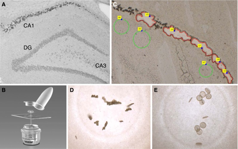
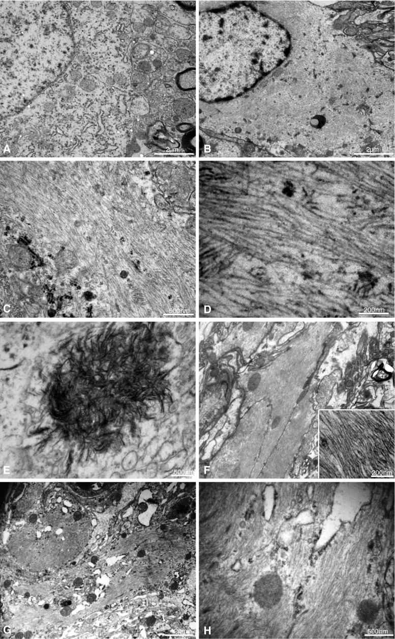
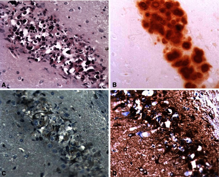
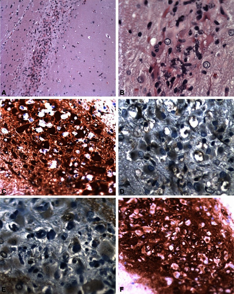
The environmental neurotoxin β-N-methylamino-L-alanine (BMAA) has been implicated in the etiology of neurodegenerative disease, and recent studies indicate that BMAA can be misincorporated into proteins. BMAA is a developmental neurotoxicant that can induce long-term learning and memory deficits, as well as regionally restricted neuronal degeneration and mineralization in the hippocampal CA1. The aim of the study was to characterize long-term changes (2 weeks to 6 months) further in the brain of adult rats treated neonatally (postnatal days 9-10) with BMAA (460 mg/kg) using immunohistochemistry (IHC), transmission electron microscopy, and laser capture microdissection followed by LC-MS/MS for proteomic analysis. The histological examination demonstrated progressive neurodegenerative changes, astrogliosis, microglial activation, and calcification in the hippocampal CA1 3-6 months after exposure. The IHC showed an increased staining for α-synuclein and ubiquitin in the area. The ultrastructural examination revealed intracellular deposition of abundant bundles of closely packed parallel fibrils in neurons, axons, and astrocytes of the CA1. Proteomic analysis of the affected site demonstrated an enrichment of chaperones (e.g., clusterin, GRP-78), cytoskeletal and intermediate filament proteins, and proteins involved in the antioxidant defense system. Several of the most enriched proteins (plectin, glial fibrillar acidic protein, vimentin, Hsp 27, and ubiquitin) are known to form complex astrocytic inclusions, so-called Rosenthal fibers, in the neurodegenerative disorder Alexander disease. In addition, TDP-43 and the negative regulator of autophagy, GLIPR-2, were exclusively detected. The present study demonstrates that neonatal exposure to BMAA may offer a novel model for the study of hippocampal fibril formation in vivo.
环境神经毒素β-N-甲基氨基-L-丙氨酸(BMAA)与神经退行性疾病的病因有关,最近的研究表明BMAA可错误掺入蛋白质中。BMAA是一种发育性神经毒物,可导致长期学习和记忆缺陷,以及海马CA1区局部性神经元变性和矿化。本研究的目的是,利用免疫组织化学(IHC)、透射电子显微镜和激光捕获显微切割技术,随后进行LC-MS/MS蛋白质组分析,进一步表征新生期(出生后第9 - 10天)用BMAA(460 mg/kg)处理的成年大鼠大脑中(2周龄至6月龄)的长期变化。组织学检查显示,暴露后3 - 6个月,海马CA1区出现进行性神经退行性变化、星形胶质细胞增生、小胶质细胞活化和钙化。免疫组织化学显示该区域α-突触核蛋白和泛素染色增加。超微结构检查显示CA1区的神经元、轴突和星形胶质细胞内有大量紧密排列的平行纤维束的细胞内沉积。对受影响部位的蛋白质组分析表明,伴侣蛋白(如簇集蛋白、GRP-78)、细胞骨架和中间丝蛋白以及参与抗氧化防御系统的蛋白富集。几种最富集的蛋白质(网蛋白、胶质纤维酸性蛋白、波形蛋白、热休克蛋白27和泛素)已知在神经退行性疾病亚历山大病中形成复杂的星形胶质细胞内含物,即所谓的罗森塔尔纤维。此外,还专门检测到了TDP-43和自噬负调节因子GLIPR-2。本研究表明,新生期暴露于BMAA可能为体内海马纤维形成的研究提供一种新模型。