Lungkaphin Anusorn, Arjinajarn Phatchawan, Pongchaidecha Anchalee, Srimaroeng Chutima, Chatsudthipong Lisa, Chatsudthipong Varanuj
Department of Physiology, Faculty of Medicine, Chiang Mai University, Chiang Mai, Thailand.
Department of Biology, Faculty of Science, Chiang Mai University, Chiang Mai, Thailand.
PLoS One. 2014 May 6;9(5):e96236. doi: 10.1371/journal.pone.0096236. eCollection 2014.
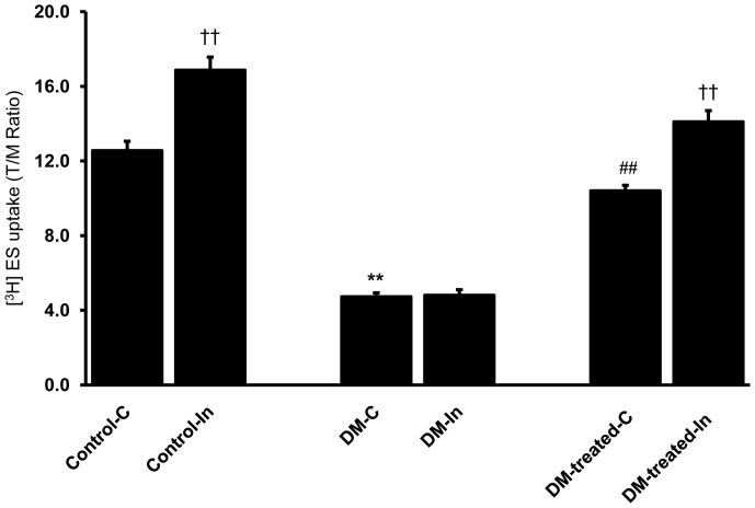
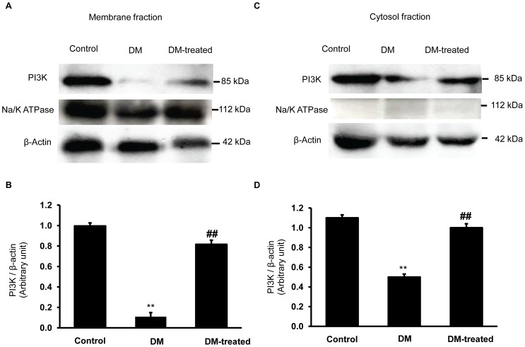
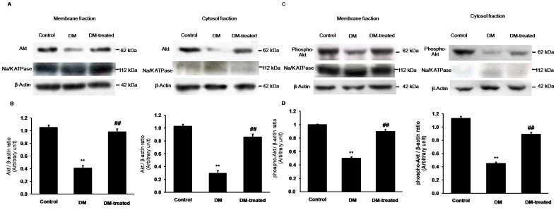
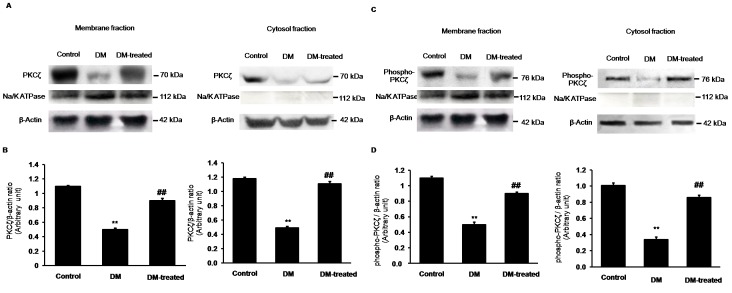
Organic anion transporter 3 (Oat3) is a major renal Oats expressed in the basolateral membrane of renal proximal tubule cells. We have recently reported decreases in renal Oat3 function and expression in diabetic rats and these changes were recovered after insulin treatment for four weeks. However, the mechanisms by which insulin restored these changes have not been elucidated. In this study, we hypothesized that insulin signaling mediators might play a crucial role in the regulation of renal Oat3 function. Experimental diabetic rats were induced by a single intraperitoneal injection of streptozotocin (65 mg/kg). One week after injection, animals showing blood glucose above 250 mg/dL were considered to be diabetic and used for the experiment in which insulin-treated diabetic rats were subcutaneously injected daily with insulin for four weeks. Estrone sulfate (ES) uptake into renal cortical slices was examined to reflect the renal Oat3 function. The results showed that pre-incubation with insulin for 30 min (short term) stimulated [3H]ES uptake into the renal cortical slices of normal control rats. In the untreated diabetic rats, pre-incubation with insulin for 30 min failed to stimulate renal Oat3 activity. The unresponsiveness of renal Oat3 activity to insulin in the untreated diabetic rats suggests the impairment of insulin signaling. Indeed, pre-incubation with phosphoinositide 3-kinase (PI3K) and protein kinase C zeta (PKCζ) inhibitors inhibited insulin-stimulated renal Oat3 activity. In addition, the expressions of PI3K, Akt and PKCζ in the renal cortex of diabetic rats were markedly decreased. Prolonged insulin treatment in diabetic rats restored these alterations toward normal levels. Our data suggest that the decreases in both function and expression of renal Oat3 in diabetes are associated with an impairment of renal insulin-induced Akt/PKB activation through PI3K/PKCζ/Akt/PKB signaling pathway.
有机阴离子转运体3(Oat3)是一种主要的肾脏有机阴离子转运体,表达于肾近端小管细胞的基底外侧膜。我们最近报道,糖尿病大鼠的肾脏Oat3功能和表达降低,胰岛素治疗四周后这些变化得以恢复。然而,胰岛素恢复这些变化的机制尚未阐明。在本研究中,我们假设胰岛素信号介质可能在肾脏Oat3功能的调节中起关键作用。通过单次腹腔注射链脲佐菌素(65mg/kg)诱导实验性糖尿病大鼠。注射一周后,血糖高于250mg/dL的动物被认为患有糖尿病,并用于胰岛素治疗的糖尿病大鼠实验,这些大鼠每天皮下注射胰岛素,持续四周。检测硫酸雌酮(ES)进入肾皮质切片的摄取情况,以反映肾脏Oat3功能。结果显示,正常对照大鼠的肾皮质切片与胰岛素预孵育30分钟(短期)可刺激[3H]ES摄取。在未经治疗的糖尿病大鼠中,与胰岛素预孵育30分钟未能刺激肾脏Oat3活性。未经治疗的糖尿病大鼠中肾脏Oat3活性对胰岛素无反应提示胰岛素信号受损。事实上,与磷酸肌醇3激酶(PI3K)和蛋白激酶Cζ(PKCζ)抑制剂预孵育可抑制胰岛素刺激的肾脏Oat3活性。此外,糖尿病大鼠肾皮质中PI3K、Akt和PKCζ的表达明显降低。糖尿病大鼠长期胰岛素治疗可使这些改变恢复至正常水平。我们的数据表明,糖尿病中肾脏Oat3功能和表达的降低与通过PI3K/PKCζ/Akt/PKB信号通路的肾脏胰岛素诱导的Akt/PKB激活受损有关。