Tan Chunlai, Lui Pauline Po Yee, Lee Yuk Wa, Wong Yin Mei
Department of Orthopaedics and Traumatology, Faculty of Medicine, The Chinese University of Hong Kong, Hong Kong SAR, China; The Hong Kong Jockey Club Sports Medicine and Health Sciences Centre, Faculty of Medicine, The Chinese University of Hong Kong, Hong Kong SAR, China.
Headquarter, Hospital Authority, Hong Kong SAR, China.
PLoS One. 2014 May 15;9(5):e97453. doi: 10.1371/journal.pone.0097453. eCollection 2014.
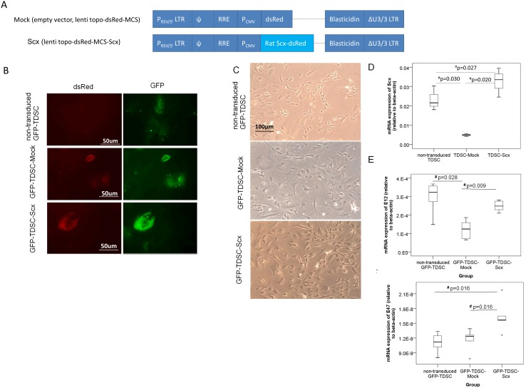
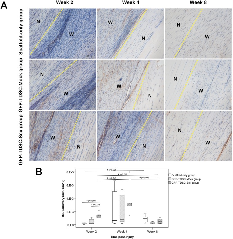
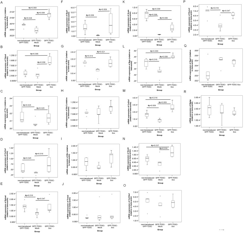
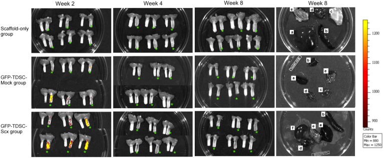
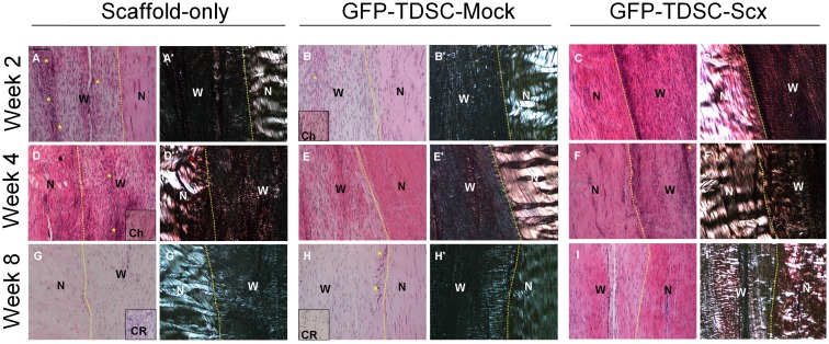
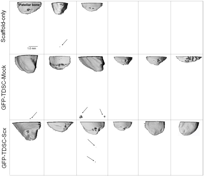
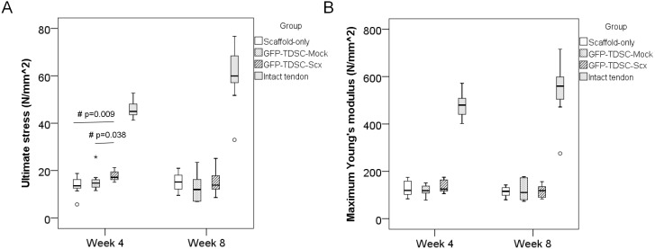
We hypothesized that the transplantation of Scx-transduced tendon-derived stem cells (TDSCs) promoted better tendon repair compared to the transplantation of mock-transduced cells. This study thus aimed to investigate the effect of Scx transduction on the expression of lineage markers in TDSCs and the effect of the resulting cell line in the promotion of tendon repair. Rat non-GFP or GFP-TDSCs were transduced with Scx or empty lentiviral vector (Mock) and selected by blasticidin. The mRNA expressions of Scx and different lineage markers were examined by qRT-PCR. The effect of the transplantation of GFP-TDSC-Scx on tendon repair was then tested in a rat unilateral patellar tendon window injury model. The transplantation of GFP-TDSC-Mock and scaffold-only served as controls. At week 2, 4 and 8 post-transplantation, the repaired patellar tendon was harvested for ex vivo fluorescent imaging, vivaCT imaging, histology, immunohistochemistry and biomechanical test. GFP-TDSC-Scx consistently showed higher expressions of most of tendon- and cartilage- related markers compared to the GFP-TDSC-Mock. However, the effect of Scx transduction on the expressions of bone-related markers was inconclusive. The transplanted GFP-TDSCs could be detected in the window wound at week 2 but not at week 4. Ectopic mineralization was detected in some samples at week 8 but there was no difference among different groups. The GFP-TDSC-Scx group only statistically significantly improved tendon repair histologically and biomechanically compared to the Scaffold-only group and the GFP-TDSC-Mock group at the early stage of tendon repair. There was significant higher expression of collagen type I in the window wound in the GFP-TDSC-Scx group compared to the other two groups at week 2. The transplantation of GFP-TDSC-Scx promoted healing at the early stage of tendon repair in a rat patellar tendon window injury model.
我们假设,与转导空载体的细胞移植相比,转导Scx的肌腱来源干细胞(TDSCs)移植能促进更好的肌腱修复。因此,本研究旨在探讨Scx转导对TDSCs中谱系标志物表达的影响,以及由此产生的细胞系在促进肌腱修复中的作用。用Scx或空慢病毒载体(Mock)转导大鼠非绿色荧光蛋白(GFP)或GFP-TDSCs,并通过杀稻瘟菌素进行筛选。通过qRT-PCR检测Scx和不同谱系标志物的mRNA表达。然后在大鼠单侧髌腱窗口损伤模型中测试GFP-TDSC-Scx移植对肌腱修复的影响。GFP-TDSC-Mock移植和仅植入支架作为对照。在移植后第2、4和8周,采集修复后的髌腱进行离体荧光成像、活体CT成像、组织学、免疫组织化学和生物力学测试。与GFP-TDSC-Mock相比,GFP-TDSC-Scx始终显示出大多数肌腱和软骨相关标志物的更高表达。然而,Scx转导对骨相关标志物表达的影响尚无定论。移植的GFP-TDSCs在第2周时可在窗口伤口中检测到,但在第4周时未检测到。在第8周时,在一些样本中检测到异位矿化,但不同组之间没有差异。在肌腱修复早期,与仅植入支架组和GFP-TDSC-Mock组相比,GFP-TDSC-Scx组在组织学和生物力学方面仅在统计学上显著改善了肌腱修复。在第2周时,与其他两组相比,GFP-TDSC-Scx组窗口伤口中I型胶原蛋白的表达显著更高。在大鼠髌腱窗口损伤模型中,GFP-TDSC-Scx移植促进了肌腱修复早期的愈合。