Institut du Cerveau et de la Moelle Épinière, Hôpital de la Pitié-Salpêtrière Paris, France ; Institut National de la Santé et de la Recherche Médicale UMR 1127 Paris, France ; Centre National de la Recherche Scientifique UMR 7225 Paris, France ; UPMC Univ. Paris 06 Paris, France ; Muséum National d'Histoire Naturelle Paris, France ; Centre National de la Recherche Scientifique UMR 7221 Paris, France.
Institut du Cerveau et de la Moelle Épinière, Hôpital de la Pitié-Salpêtrière Paris, France ; Institut National de la Santé et de la Recherche Médicale UMR 1127 Paris, France ; Centre National de la Recherche Scientifique UMR 7225 Paris, France ; UPMC Univ. Paris 06 Paris, France.
Front Neuroanat. 2014 May 6;8:26. doi: 10.3389/fnana.2014.00026. eCollection 2014.
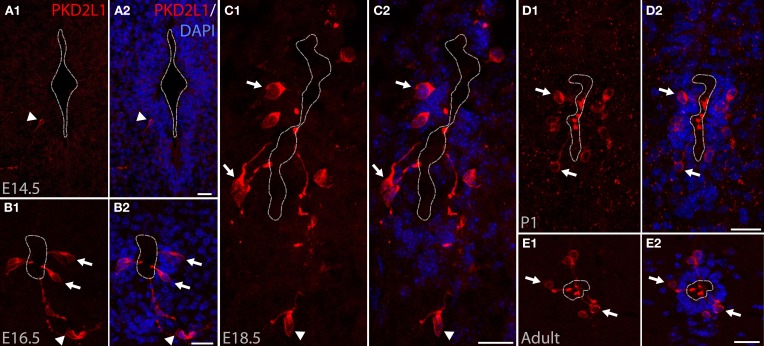
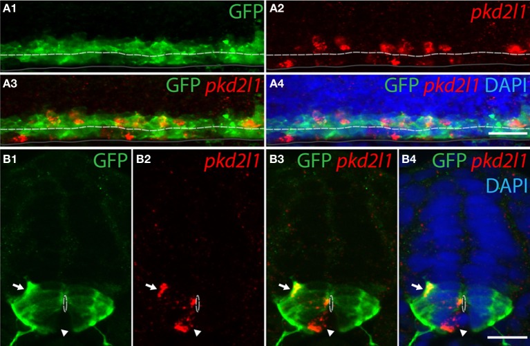
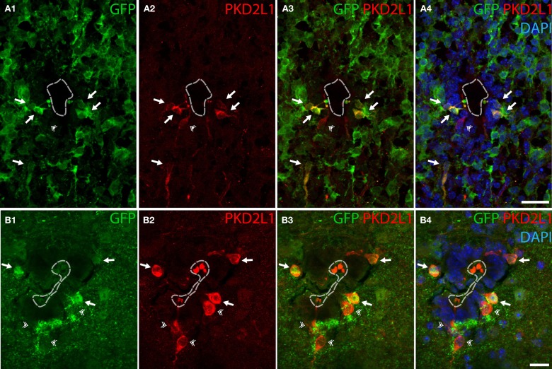
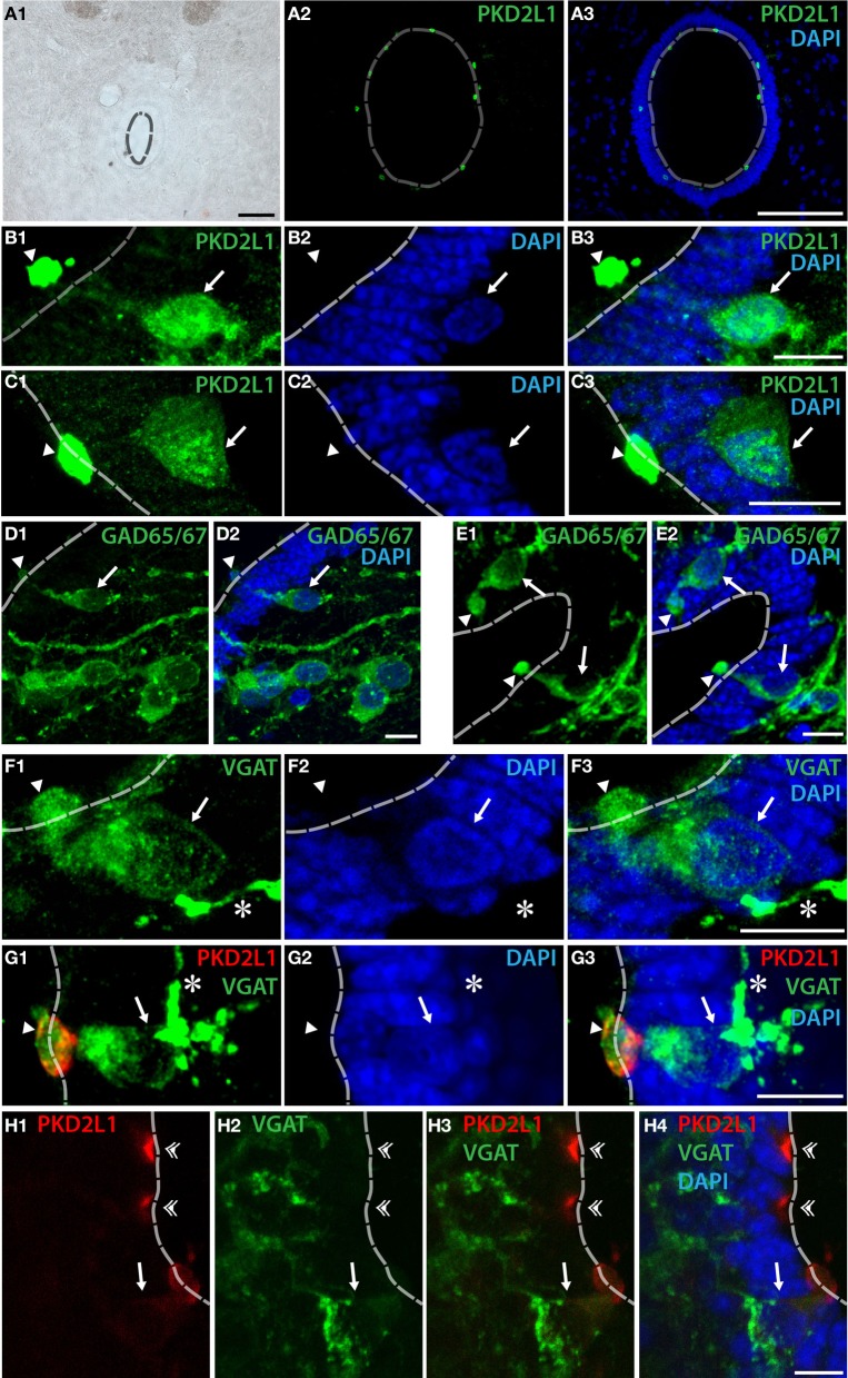
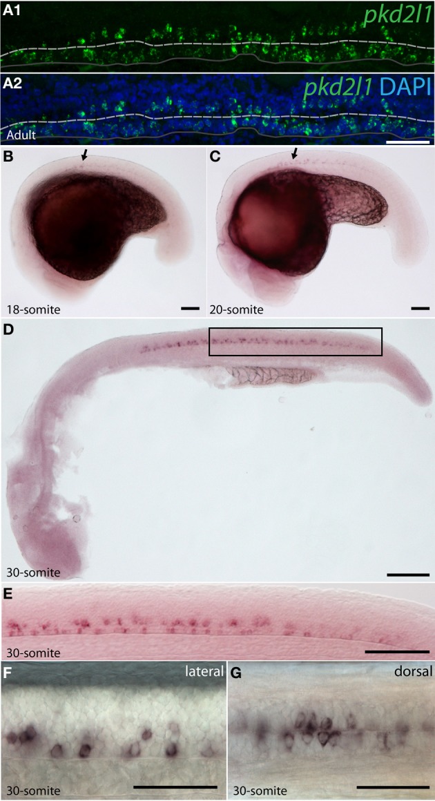
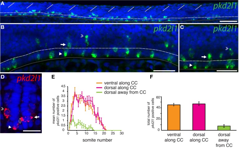
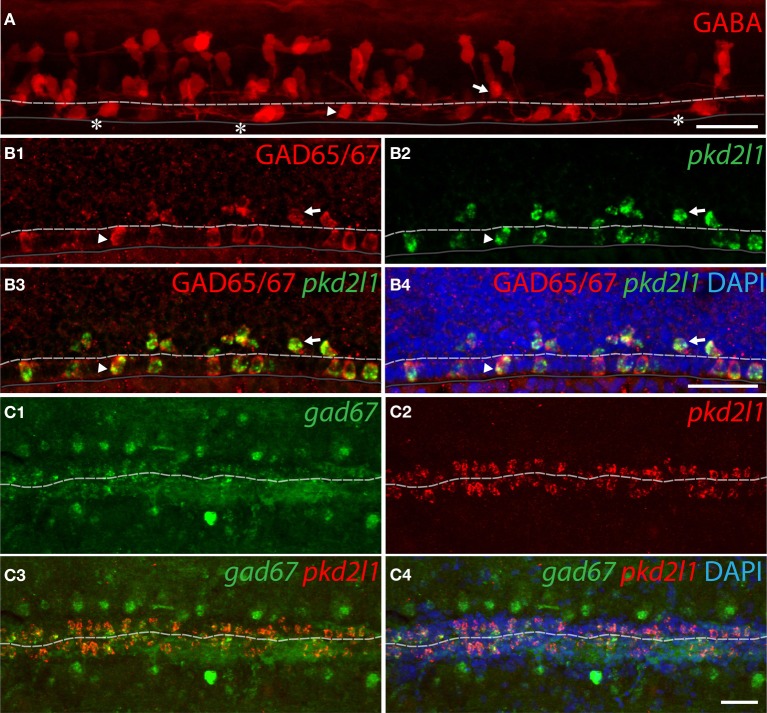
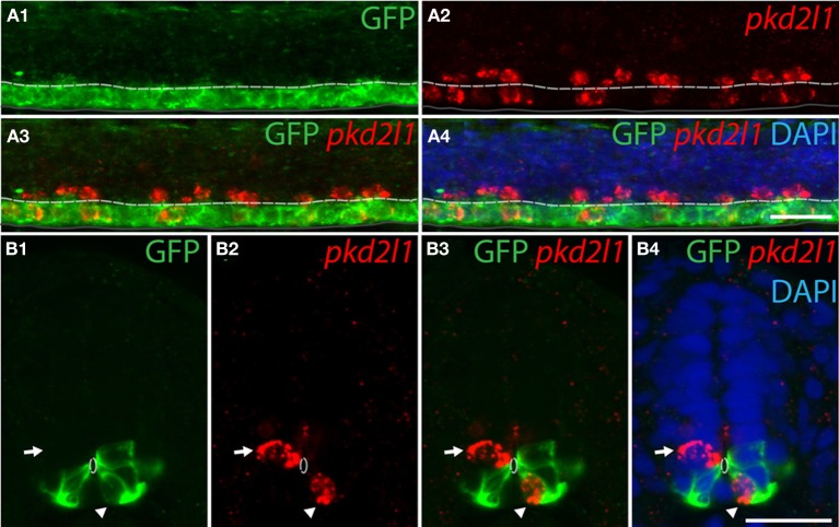
Over 90 years ago, Kolmer and Agduhr identified spinal cerebrospinal fluid-contacting neurons (CSF-cNs) based on their morphology and location within the spinal cord. In more than 200 vertebrate species, they observed ciliated neurons around the central canal that extended a brush of microvilli into the cerebrospinal fluid (CSF). Although their morphology is suggestive of a primitive sensory cell, their function within the vertebrate spinal cord remains unknown. The identification of specific molecular markers for these neurons in vertebrates would benefit the investigation of their physiological roles. PKD2L1, a transient receptor potential channel that could play a role as a sensory receptor, has been found in cells contacting the central canal in mouse. In this study, we demonstrate that PKD2L1 is a specific marker for CSF-cNs in the spinal cord of mouse (Mus musculus), macaque (Macaca fascicularis) and zebrafish (Danio rerio). In these species, the somata of spinal PKD2L1(+) CSF-cNs were located below or within the ependymal layer and extended an apical bulbous extension into the central canal. We found GABAergic PKD2L1-expressing CSF-cNs in all three species. We took advantage of the zebrafish embryo for its transparency and rapid development to identify the progenitor domains from which pkd2l1 (+) CSF-cNs originate. pkd2l1 (+) CSF-cNs were all GABAergic and organized in two rows-one ventral and one dorsal to the central canal. Their location and marker expression is consistent with previously described Kolmer-Agduhr cells. Accordingly, pkd2l1 (+) CSF-cNs were derived from the progenitor domains p3 and pMN defined by the expression of nkx2.2a and olig2 transcription factors, respectively. Altogether our results suggest that a system of CSF-cNs expressing the PKD2L1 channel is conserved in the spinal cord across bony vertebrate species.
90 多年前,Kolmer 和 Agduhr 根据形态学和脊髓内位置确定了脊髓脑脊液接触神经元 (CSF-cNs)。在 200 多种脊椎动物中,他们观察到中央管周围有纤毛神经元,这些神经元伸出一束微绒毛进入脑脊液 (CSF)。尽管它们的形态暗示了原始感觉细胞,但它们在脊椎动物脊髓中的功能仍然未知。在脊椎动物中鉴定这些神经元的特定分子标记将有助于研究它们的生理作用。PKD2L1 是一种瞬时受体电位通道,可能作为感觉受体发挥作用,已在小鼠接触中央管的细胞中发现。在这项研究中,我们证明 PKD2L1 是小鼠 (Mus musculus)、猕猴 (Macaca fascicularis) 和斑马鱼 (Danio rerio) 脊髓 CSF-cNs 的特异性标记物。在这些物种中,脊髓 PKD2L1(+) CSF-cNs 的体位于室管膜层下方或内部,并向中央管延伸一个顶端球状延伸。我们在所有三个物种中都发现了 GABA 能 PKD2L1 表达的 CSF-cNs。我们利用斑马鱼胚胎的透明性和快速发育来鉴定起源于 pkd2l1 (+) CSF-cNs 的祖细胞域。pkd2l1 (+) CSF-cNs 均为 GABA 能神经元,排列在中央管的腹侧和背侧各一行。它们的位置和标记物表达与先前描述的 Kolmer-Agduhr 细胞一致。因此,pkd2l1 (+) CSF-cNs 分别由 nkxa2.2a 和 olig2 转录因子表达定义的祖细胞域 p3 和 pMN 衍生而来。总的来说,我们的结果表明,在整个硬骨脊椎动物物种的脊髓中,表达 PKD2L1 通道的 CSF-cNs 系统是保守的。