Nofiele Joris Tchouala, Cheng Hai-Ling Margaret
The Research Institute and Diagnostic Imaging, Hospital for Sick Children, Toronto, Ontario, Canada; Department of Medical Biophysics, University of Toronto, Toronto, Ontario, Canada.
The Research Institute and Diagnostic Imaging, Hospital for Sick Children, Toronto, Ontario, Canada; Department of Medical Biophysics, University of Toronto, Toronto, Ontario, Canada; Leslie Dan Faculty of Pharmacy, University of Toronto, Toronto, Ontario, Canada; The Institute of Biomaterials & Biomedical Engineering, University of Toronto, Toronto, Ontario, Canada.
PLoS One. 2014 May 16;9(5):e97950. doi: 10.1371/journal.pone.0097950. eCollection 2014.
Larger animal models provide relevant tumor burden in the development of advanced clinical imaging methods for non-invasive cancer detection and diagnosis, and are especially valuable for studying metastatic disease. Most available experimental models, however, are based on immune-compromised mice. To lay the foundation for studying spontaneous metastasis using non-invasive magnetic resonance imaging (MRI), this study aims to establish a highly metastatic breast cancer xenograft model in nude rats.
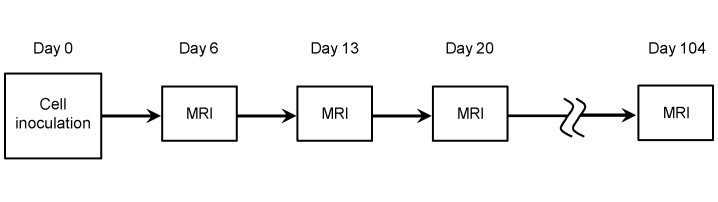
A highly metastatic variant of human adenocarcinoma MDA-MB-231 known as LM2 was inoculated into nude rats. Orthotopic and subcutaneous (flank) sites were compared, with half of the orthotopic injections guided by ultrasound imaging. MRI with gadolinium contrast administration was performed weekly beginning on Day 6 and ending on Day 104. Excised tumors were assessed on histology using hematoxylin and eosin and CD34. Fisher's exact test was used to compare successful tumor induction amongst different inoculation methods.
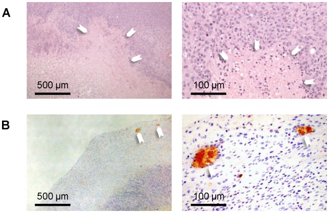
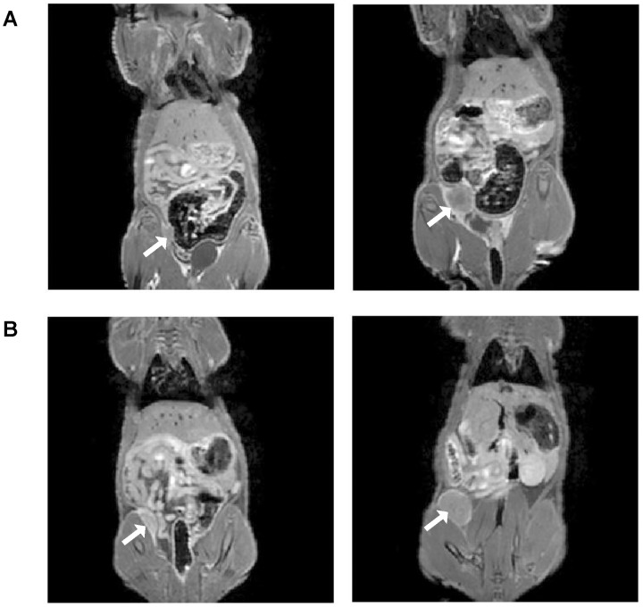
Primary LM2 tumors were established orthotopically in all cases under ultrasound-guided injection, and none otherwise (p = 0.0028). Contrast-enhanced MRI revealed rapidly progressing tumors that reached critical size (15 mm diameter) in 2 to 3 weeks after inoculation. MRI and histology findings were consistent: LM2 tumors were characterized by low vascularity confined to the tumor rim and large necrotic cores with increasing interstitial fluid pressure.
The metastatic LM2 breast tumor model was successfully established in the mammary fat pads of nude rats, using ultrasound needle guidance as a non-invasive alternative to surgery. This platform lays the foundation for future development and application of MRI to study spontaneous metastasis and different stages throughout the metastatic cascade.
在开发用于非侵入性癌症检测和诊断的先进临床成像方法时,大型动物模型能提供相关的肿瘤负荷,对研究转移性疾病尤其有价值。然而,现有的大多数实验模型都基于免疫缺陷小鼠。为了为使用非侵入性磁共振成像(MRI)研究自发性转移奠定基础,本研究旨在建立一种裸鼠高转移性乳腺癌异种移植模型。
将一种已知的人腺癌MDA-MB-231的高转移性变体LM2接种到裸鼠体内。比较了原位和皮下(侧腹)接种部位,其中一半原位注射在超声成像引导下进行。从第6天开始至第104天结束,每周进行一次钆对比剂增强MRI检查。使用苏木精和伊红以及CD34对切除的肿瘤进行组织学评估。采用Fisher精确检验比较不同接种方法的肿瘤诱导成功率。
在超声引导注射下,所有病例均成功原位建立了原发性LM2肿瘤,否则无一成功(p = 0.0028)。对比增强MRI显示肿瘤进展迅速,接种后2至3周达到临界大小(直径15 mm)。MRI和组织学结果一致:LM2肿瘤的特征是血管分布少,局限于肿瘤边缘,坏死核心大,间质液压力增加。
利用超声针引导作为手术的非侵入性替代方法,在裸鼠的乳腺脂肪垫中成功建立了转移性LM2乳腺肿瘤模型。该平台为未来开发和应用MRI研究自发性转移以及转移级联反应的不同阶段奠定了基础。