Birn R M, Shackman A J, Oler J A, Williams L E, McFarlin D R, Rogers G M, Shelton S E, Alexander A L, Pine D S, Slattery M J, Davidson R J, Fox A S, Kalin N H
1] Department of Medical Physics, University of Wisconsin, Madison, WI, USA [2] Department of Psychiatry, University of Wisconsin, Madison, WI, USA [3] HealthEmotions Research Institute, Wisconsin Psychiatric Institute and Clinics, University of Wisconsin, Madison, WI, USA [4] Lane Neuroimaging Laboratory, University of Wisconsin, Madison, WI, USA [5] Waisman Laboratory for Brain Imaging and Behavior, University of Wisconsin, Madison, WI, USA.
1] Department of Psychology, University of Maryland, College Park, MD, USA [2] Neuroscience and Cognitive Science Program, University of Maryland, College Park, MD, USA [3] Maryland Neuroimaging Center, University of Maryland, College Park, MD, USA.
Mol Psychiatry. 2014 Aug;19(8):915-22. doi: 10.1038/mp.2014.46. Epub 2014 May 27.
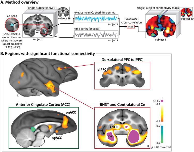
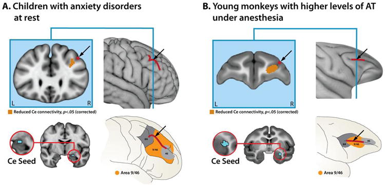
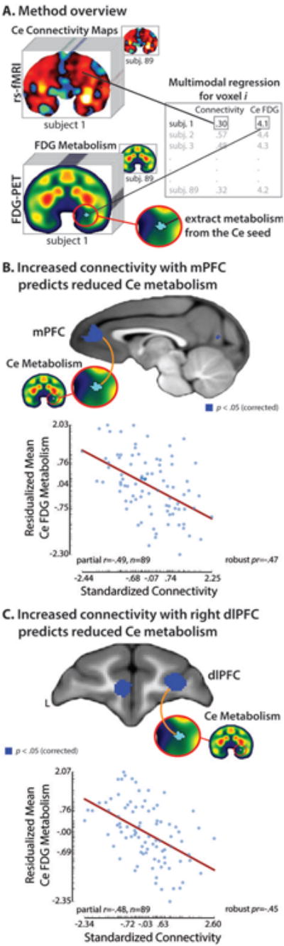
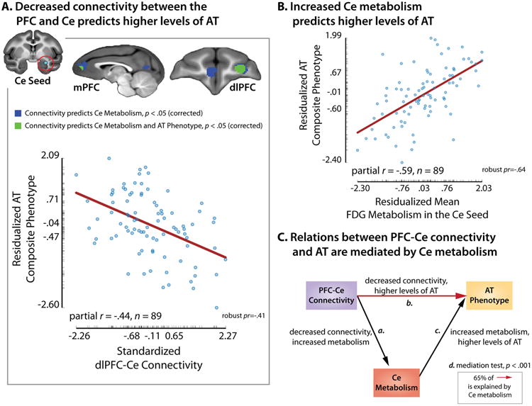
Some individuals are endowed with a biology that renders them more reactive to novelty and potential threat. When extreme, this anxious temperament (AT) confers elevated risk for the development of anxiety, depression and substance abuse. These disorders are highly prevalent, debilitating and can be challenging to treat. The high-risk AT phenotype is expressed similarly in children and young monkeys and mechanistic work demonstrates that the central (Ce) nucleus of the amygdala is an important substrate. Although it is widely believed that the flow of information across the structural network connecting the Ce nucleus to other brain regions underlies primates' capacity for flexibly regulating anxiety, the functional architecture of this network has remained poorly understood. Here we used functional magnetic resonance imaging (fMRI) in anesthetized young monkeys and quietly resting children with anxiety disorders to identify an evolutionarily conserved pattern of functional connectivity relevant to early-life anxiety. Across primate species and levels of awareness, reduced functional connectivity between the dorsolateral prefrontal cortex, a region thought to play a central role in the control of cognition and emotion, and the Ce nucleus was associated with increased anxiety assessed outside the scanner. Importantly, high-resolution 18-fluorodeoxyglucose positron emission tomography imaging provided evidence that elevated Ce nucleus metabolism statistically mediates the association between prefrontal-amygdalar connectivity and elevated anxiety. These results provide new clues about the brain network underlying extreme early-life anxiety and set the stage for mechanistic work aimed at developing improved interventions for pediatric anxiety.
一些个体天生具有一种生物学特性,使他们对新事物和潜在威胁反应更强烈。当这种情况极端化时,这种焦虑气质(AT)会增加患焦虑症、抑郁症和药物滥用的风险。这些疾病非常普遍,使人衰弱,且治疗颇具挑战性。高风险的AT表型在儿童和幼年猴子中表现相似,机理研究表明杏仁核的中央(Ce)核是一个重要基础。尽管人们普遍认为,信息在连接Ce核与其他脑区的结构网络中的流动是灵长类动物灵活调节焦虑能力的基础,但这个网络的功能架构仍知之甚少。在这里,我们对麻醉状态下的幼年猴子和安静休息的焦虑症儿童进行了功能磁共振成像(fMRI),以识别与早期生活焦虑相关的进化保守的功能连接模式。在灵长类物种和意识水平中,背外侧前额叶皮层(一个被认为在认知和情绪控制中起核心作用的区域)与Ce核之间功能连接的减少,与扫描仪外评估的焦虑增加有关。重要的是,高分辨率18-氟脱氧葡萄糖正电子发射断层扫描成像提供了证据,表明Ce核代谢升高在统计学上介导了前额叶-杏仁核连接与焦虑升高之间的关联。这些结果为极端早期生活焦虑背后的脑网络提供了新线索,并为旨在开发改善儿童焦虑症干预措施的机理研究奠定了基础。