• 文献检索
  • 文档翻译
  • 深度研究
  • 学术资讯
  • Suppr Zotero 插件Zotero 插件
  • 邀请有礼
  • 套餐&价格
  • 历史记录
应用&插件
Suppr Zotero 插件Zotero 插件浏览器插件Mac 客户端Windows 客户端微信小程序
定价
高级版会员购买积分包购买API积分包
服务
文献检索文档翻译深度研究API 文档MCP 服务
关于我们
关于 Suppr公司介绍联系我们用户协议隐私条款
关注我们

Suppr 超能文献

核心技术专利:CN118964589B侵权必究
粤ICP备2023148730 号-1Suppr @ 2026

文献检索

告别复杂PubMed语法,用中文像聊天一样搜索,搜遍4000万医学文献。AI智能推荐,让科研检索更轻松。

立即免费搜索

文件翻译

保留排版,准确专业,支持PDF/Word/PPT等文件格式,支持 12+语言互译。

免费翻译文档

深度研究

AI帮你快速写综述,25分钟生成高质量综述,智能提取关键信息,辅助科研写作。

立即免费体验

壳核低信号在帕金森病型多系统萎缩中的表现:应用磁敏感加权成像的简单视觉评估。

Putaminal hypointensity in the parkinsonian variant of multiple system atrophy: simple visual assessment using susceptibility-weighted imaging.

机构信息

Department of Neurology, Research Institute for Convergence of Biomedical Science and Technology, Pusan National University Yangsan Hospital, Yangsan, Korea.

Department of Diagnostic Radiology, Research Institute for Convergence of Biomedical Science and Technology, Pusan National University Yangsan Hospital, Yangsan, Korea.

出版信息

J Mov Disord. 2011 Oct;4(2):60-3. doi: 10.14802/jmd.11012. Epub 2011 Oct 30.

DOI:10.14802/jmd.11012
PMID:24868396
原文链接:https://pmc.ncbi.nlm.nih.gov/articles/PMC4027686/
Abstract

BACKGROUND AND PURPOSE

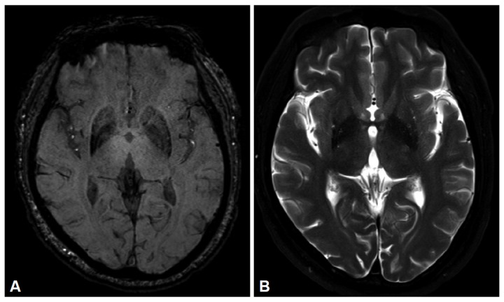
Susceptibility-weighted imaging (SWI) has been shown to be superior in its ability to demonstrate brain mineralization than other conventional MR imaging. The goal of our study was therefore to assess the frequency and extent of putaminal hypointensity in parkinsonian variant MSA using SWI.

METHODS

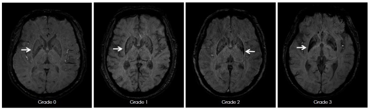
11 patients with multiple system atrophy-parkinsonian type (MSA-p), 30 patients with Parkinson's disease (PD), and age matched 30 controls were investigated using 3 Tesla MRI. The pattern of putaminal hypointensity was measured using a visual grading scale and scored from 0 to 3.

RESULTS

Hemi- or bilateral putaminal hypointensity (a score of ≥ 2) and hyperintense rim were recognized in 81.8% and 54.5% of 11 MSA-p, respectively. The scores of putaminal hypointensity of MSA-p were significantly higher than other groups (p < 0.001), a score of ≥ 2 differentiated MSA-p from other groups. And all five patients with early disease stage also showed these characteristic findings.

CONCLUSIONS

SWI appears to be useful for depicting putaminal hypointensity even in early stage of MSA-p. This finding suggests that iron deposition associated putaminal degeneration can occur early in the disease process.

摘要

背景与目的

磁共振磁敏感加权成像(SWI)在显示脑矿化方面优于其他常规磁共振成像。因此,我们的研究目的是使用 SWI 评估帕金森变异型多系统萎缩(MSA-p)患者壳核低信号的频率和程度。

方法

使用 3.0T MRI 对 11 例多系统萎缩-帕金森型(MSA-p)患者、30 例帕金森病(PD)患者和年龄匹配的 30 例对照组进行研究。采用视觉评分量表测量壳核低信号的模式,并评分 0-3 分。

结果

81.8%和 54.5%的 11 例 MSA-p 患者分别存在半侧或双侧壳核低信号(评分≥2)和高信号环。MSA-p 患者的壳核低信号评分明显高于其他组(p<0.001),评分≥2 可将 MSA-p 与其他组区分开来。而且,所有 5 例早期疾病阶段的患者也表现出这些特征性发现。

结论

SWI 似乎可用于描绘壳核低信号,甚至在 MSA-p 的早期阶段也如此。这一发现表明,铁沉积相关的壳核变性可能在疾病过程的早期发生。

https://cdn.ncbi.nlm.nih.gov/pmc/blobs/3e54/4027686/f25eb93749bb/jmd-4-2-60-2f2.jpg
https://cdn.ncbi.nlm.nih.gov/pmc/blobs/3e54/4027686/ee32b1251a4b/jmd-4-2-60-2f1.jpg
https://cdn.ncbi.nlm.nih.gov/pmc/blobs/3e54/4027686/f25eb93749bb/jmd-4-2-60-2f2.jpg
https://cdn.ncbi.nlm.nih.gov/pmc/blobs/3e54/4027686/ee32b1251a4b/jmd-4-2-60-2f1.jpg
https://cdn.ncbi.nlm.nih.gov/pmc/blobs/3e54/4027686/f25eb93749bb/jmd-4-2-60-2f2.jpg

相似文献

1
Putaminal hypointensity in the parkinsonian variant of multiple system atrophy: simple visual assessment using susceptibility-weighted imaging.壳核低信号在帕金森病型多系统萎缩中的表现:应用磁敏感加权成像的简单视觉评估。
J Mov Disord. 2011 Oct;4(2):60-3. doi: 10.14802/jmd.11012. Epub 2011 Oct 30.
2
Usefulness of 3D-PRESTO imaging in evaluating putaminal abnormality in parkinsonian variant of multiple system atrophy.3D-PRESTO 成像在评估帕金森病型多系统萎缩纹状体异常中的作用。
Neuroradiology. 2010 Sep;52(9):809-14. doi: 10.1007/s00234-009-0621-9. Epub 2009 Nov 6.
3
The putaminal abnormalities on 3.0T magnetic resonance imaging: can they separate parkinsonism-predominant multiple system atrophy from Parkinson's disease?3.0T磁共振成像上的壳核异常:它们能否区分以帕金森综合征为主的多系统萎缩与帕金森病?
Acta Radiol. 2015 Mar;56(3):322-8. doi: 10.1177/0284185114524090. Epub 2014 Mar 11.
4
Using 'swallow-tail' sign and putaminal hypointensity as biomarkers to distinguish multiple system atrophy from idiopathic Parkinson's disease: A susceptibility-weighted imaging study.使用“燕尾征”和壳核低信号作为生物标志物区分多系统萎缩与特发性帕金森病:一项磁敏感加权成像研究
Eur Radiol. 2017 Aug;27(8):3174-3180. doi: 10.1007/s00330-017-4743-x. Epub 2017 Jan 19.
5
Putaminal hyperintensity on T1-weighted MR imaging in patients with the Parkinson variant of multiple system atrophy.多系统萎缩帕金森变异型患者T1加权磁共振成像上的壳核高信号。
AJNR Am J Neuroradiol. 2009 Apr;30(4):689-92. doi: 10.3174/ajnr.A1443. Epub 2009 Jan 15.
6
Utility of susceptibility-weighted MRI in differentiating Parkinson's disease and atypical parkinsonism.磁共振磁敏感加权成像在鉴别帕金森病与非典型帕金森综合征中的作用。
Neuroradiology. 2010 Dec;52(12):1087-94. doi: 10.1007/s00234-010-0677-6. Epub 2010 Apr 1.
7
The "Black Straight-Line Sign" in the Putamen in Diffusion-Weighted Imaging: A Potential Diagnostic MRI Marker for Multiple System Atrophy.壳核扩散加权成像中的“黑色直线征”:多系统萎缩的一种潜在MRI诊断标志物
Front Neurol. 2022 May 19;13:890168. doi: 10.3389/fneur.2022.890168. eCollection 2022.
8
Progression of putaminal degeneration in multiple system atrophy: a serial diffusion MR study.多系统萎缩中壳核变性的进展:一项系列扩散磁共振研究。
Neuroimage. 2006 May 15;31(1):240-5. doi: 10.1016/j.neuroimage.2005.12.006. Epub 2006 Jan 27.
9
Multiparametric MR imaging of Parkinsonisms at 3 tesla: Its role in the differentiation of idiopathic Parkinson's disease versus atypical Parkinsonian disorders.3T 多参数磁共振成像在帕金森病中的应用:对特发性帕金森病与非典型帕金森综合征的鉴别诊断作用。
Eur J Radiol. 2018 Dec;109:95-100. doi: 10.1016/j.ejrad.2018.10.032. Epub 2018 Nov 1.
10
An magnetic resonance imaging T2*-weighted sequence at short echo time to detect putaminal hypointensity in Parkinsonisms.一种磁共振 T2*-加权短回波时间序列,用于检测帕金森病患者壳核的低信号。
Mov Disord. 2010 Dec 15;25(16):2728-34. doi: 10.1002/mds.23173.

引用本文的文献

1
The Irony of Iron: The Element with Diverse Influence on Neurodegenerative Diseases.铁的讽刺:对神经退行性疾病有多种影响的元素
Int J Mol Sci. 2024 Apr 12;25(8):4269. doi: 10.3390/ijms25084269.
2
Multi-parametric radiomics of conventional T1 weighted and susceptibility-weighted imaging for differential diagnosis of idiopathic Parkinson's disease and multiple system atrophy.常规 T1 加权和磁化率加权成像的多参数放射组学在特发性帕金森病和多系统萎缩鉴别诊断中的应用。
BMC Med Imaging. 2023 Dec 8;23(1):204. doi: 10.1186/s12880-023-01169-1.
3
Deep learning segmentation results in precise delineation of the putamen in multiple system atrophy.

本文引用的文献

1
Putaminal abnormality on 3-T magnetic resonance imaging in early parkinsonism-predominant multiple system atrophy.在早期帕金森病为主的多系统萎缩症的 3-T 磁共振成像上的豆状核异常。
J Neurol. 2010 Dec;257(12):2065-70. doi: 10.1007/s00415-010-5661-x. Epub 2010 Jul 22.
2
Utility of susceptibility-weighted MRI in differentiating Parkinson's disease and atypical parkinsonism.磁共振磁敏感加权成像在鉴别帕金森病与非典型帕金森综合征中的作用。
Neuroradiology. 2010 Dec;52(12):1087-94. doi: 10.1007/s00234-010-0677-6. Epub 2010 Apr 1.
3
Brain magnetic resonance imaging (MRI) in parkinsonian disorders.
深度学习分割结果精确描绘了多系统萎缩中的壳核。
Eur Radiol. 2023 Oct;33(10):7160-7167. doi: 10.1007/s00330-023-09665-2. Epub 2023 May 1.
4
The "Black Straight-Line Sign" in the Putamen in Diffusion-Weighted Imaging: A Potential Diagnostic MRI Marker for Multiple System Atrophy.壳核扩散加权成像中的“黑色直线征”:多系统萎缩的一种潜在MRI诊断标志物
Front Neurol. 2022 May 19;13:890168. doi: 10.3389/fneur.2022.890168. eCollection 2022.
5
Automated Differentiation of Atypical Parkinsonian Syndromes Using Brain Iron Patterns in Susceptibility Weighted Imaging.利用磁敏感加权成像中的脑铁模式对非典型帕金森综合征进行自动鉴别
Diagnostics (Basel). 2022 Mar 5;12(3):637. doi: 10.3390/diagnostics12030637.
6
An Updated Overview of the Magnetic Resonance Imaging of Brain Iron in Movement Disorders.运动障碍性脑铁磁共振成像的最新综述。
Behav Neurol. 2022 Feb 24;2022:3972173. doi: 10.1155/2022/3972173. eCollection 2022.
7
Diagnostic performance of T2* gradient echo, susceptibility-weighted imaging, and quantitative susceptibility mapping for patients with multiple system atrophy-parkinsonian type: a systematic review and meta-analysis.T2* 梯度回波、磁敏感加权成像和定量磁敏感图对多系统萎缩-帕金森型患者的诊断性能:系统评价和荟萃分析。
Eur Radiol. 2022 Jan;32(1):308-318. doi: 10.1007/s00330-021-08174-4. Epub 2021 Jul 16.
8
Current Management and Emerging Therapies in Multiple System Atrophy.多种系统萎缩的当前治疗和新兴疗法。
Neurotherapeutics. 2020 Oct;17(4):1582-1602. doi: 10.1007/s13311-020-00890-x.
9
Quantitative Validation of a Visual Rating Scale for Defining High-Iron Putamen in Patients With Multiple System Atrophy.用于定义多系统萎缩患者高铁含量壳核的视觉评分量表的定量验证
Front Neurol. 2019 Sep 20;10:1014. doi: 10.3389/fneur.2019.01014. eCollection 2019.
10
Brain Iron Accumulation in Atypical Parkinsonian Syndromes: MRI Evidences for Distinctive Patterns.非典型帕金森综合征中的脑铁蓄积:不同模式的MRI证据
Front Neurol. 2019 Feb 12;10:74. doi: 10.3389/fneur.2019.00074. eCollection 2019.
帕金森病相关疾病的脑磁共振成像(MRI)
Parkinsonism Relat Disord. 2009 Mar;15(3):165-74. doi: 10.1016/j.parkreldis.2008.04.033. Epub 2008 Dec 6.
4
Susceptibility-weighted imaging: technical aspects and clinical applications, part 1.磁敏感加权成像:技术要点与临床应用,第1部分
AJNR Am J Neuroradiol. 2009 Jan;30(1):19-30. doi: 10.3174/ajnr.A1400. Epub 2008 Nov 27.
5
Putaminal lesion in multiple system atrophy: postmortem MR-pathological correlations.多系统萎缩中的壳核病变:尸检磁共振成像与病理的相关性
Neuroradiology. 2008 Jul;50(7):559-67. doi: 10.1007/s00234-008-0381-y. Epub 2008 May 8.
6
Hyperintense putaminal rim at 3T reflects fewer ferritin deposits in the lateral marginal area of the putamen.3T时壳核边缘高信号反映壳核外侧边缘区域铁蛋白沉积较少。
AJNR Am J Neuroradiol. 2007 Apr;28(4):777-81.
7
T2*-weighted MRI in diagnosis of multiple system atrophy. A practical approach for clinicians.
J Neurol. 2007 Sep;254(9):1184-8. doi: 10.1007/s00415-006-0496-1. Epub 2007 Mar 14.
8
Evaluating posterolateral linearization of the putaminal margin with magnetic resonance imaging to diagnose the Parkinson variant of multiple system atrophy.利用磁共振成像评估壳核边缘的后外侧线性化以诊断多系统萎缩的帕金森变异型。
Mov Disord. 2007 Mar 15;22(4):578-81. doi: 10.1002/mds.21329.
9
Hyperintense putaminal rim sign is not a hallmark of multiple system atrophy at 3T.壳核高信号边缘征并非3T磁共振成像下多系统萎缩的特征性表现。
AJNR Am J Neuroradiol. 2005 Oct;26(9):2238-42.
10
Comparison of magnetic resonance imaging in subtypes of multiple system atrophy.多系统萎缩各亚型的磁共振成像比较
Parkinsonism Relat Disord. 2004 Aug;10(6):363-8. doi: 10.1016/j.parkreldis.2004.04.008.