The Center of Research Laboratory, and Department of Gynecology, The International Peace Maternity and Child Health Hospital, School of Medicine, Shanghai Jiaotong University, Shanghai, China.
PLoS One. 2014 May 30;9(5):e98749. doi: 10.1371/journal.pone.0098749. eCollection 2014.
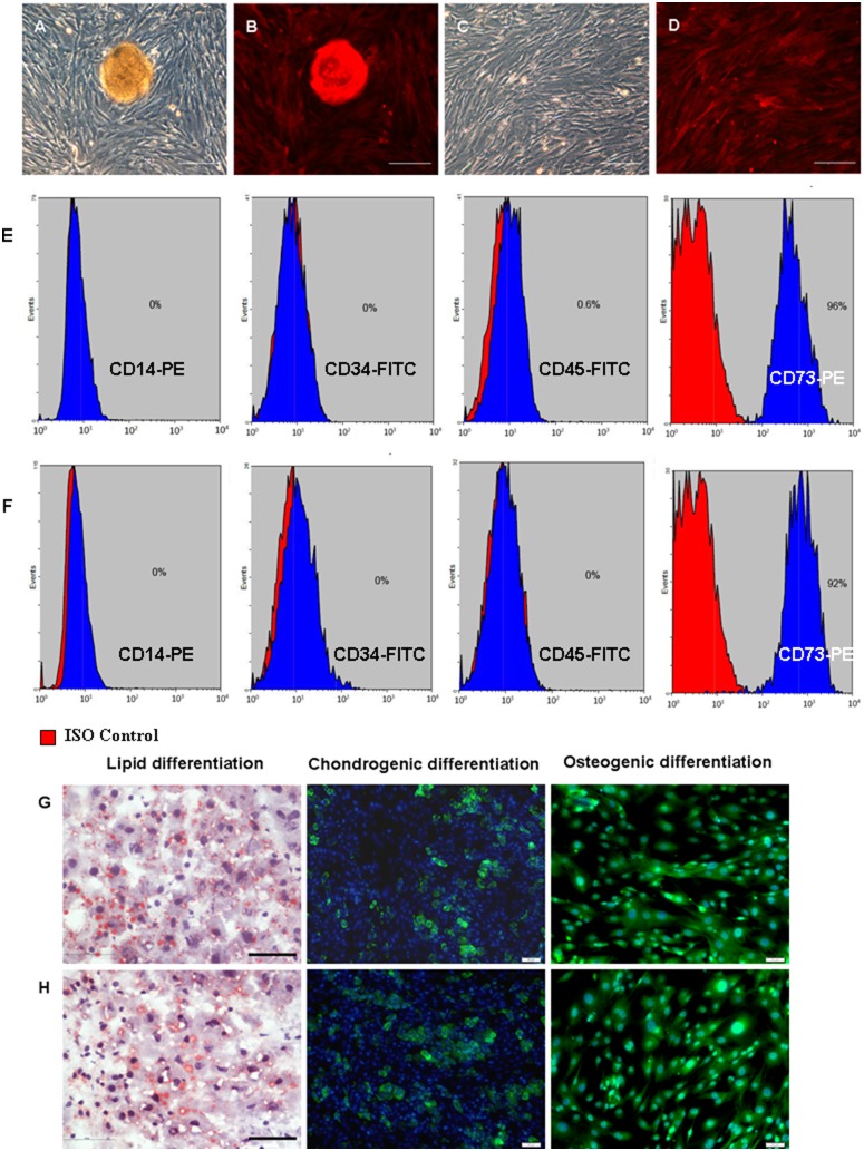
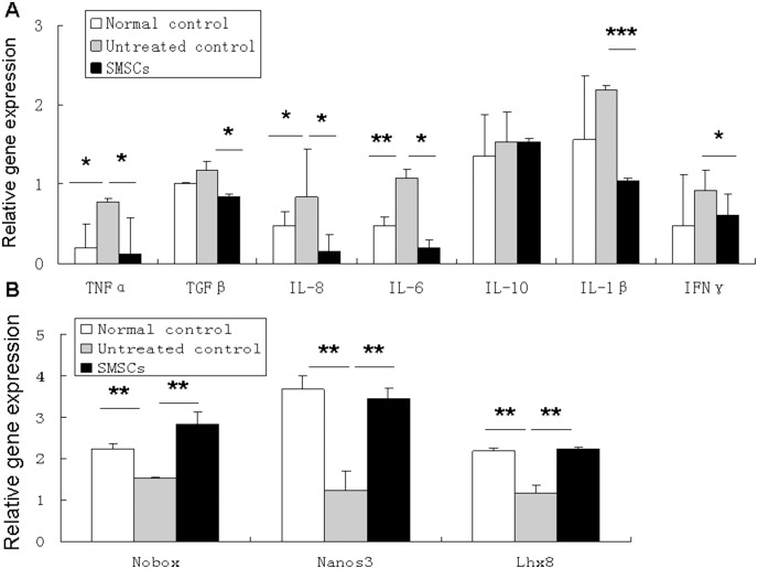
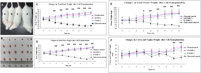
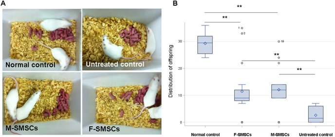
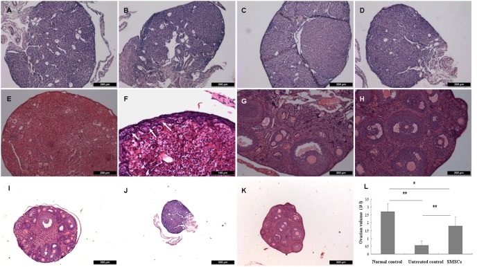
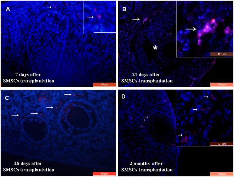
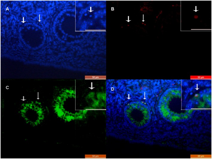
Skin-derived mesenchymal stem cells (SMSCs) can differentiate into the three embryonic germ layers. For this reason, they are considered a powerful tool for therapeutic cloning and offer new possibilities for tissue therapy. Recent studies showed that skin-derived stem cells can differentiate into cells expressing germ-cell specific markers in vitro and form oocytes in vivo. The idea that SMSCs may be suitable for the treatment of intractable diseases or traumatic tissue damage has attracted attention. To determine the ability of SMSCs to reactivate injured ovaries, a mouse model with ovaries damaged by busulfan and cyclophosphamide was developed and is described here. Female skin-derived mesenchymal stem cells (F-SMSCs) and male skin-derived mesenchymal stem cells (M-SMSCs) from red fluorescence protein (RFP) transgenic adult mice were used to investigate the restorative effects of SMSCs on ovarian function. Significant increases in total body weight and the weight of reproductive organs were observed in the treated animals. Both F-SMSCs and M-SMSCs were shown to be capable of partially restoring fertility in chemotherapy-treated females. Immunostaining with RFP and anti-Müllerian hormone (AMH) antibodies demonstrated that the grafted SMSCs survived, migrated to the recipient ovaries. After SMSCs were administered to the treated mice, real-time PCR showed that the expression levels of pro-inflammatory cytokines TNF-α, TGF-β, IL-8, IL-6, IL-1β, and IFNγ were significantly lower in the ovaries than in the untreated controls. Consistent with this observation, expression of oogenesis marker genes Nobox, Nanos3, and Lhx8 increased in ovaries of SMSCs-treated mice. These findings suggest that SMSCs may play a role within the ovarian follicle microenvironment in restoring the function of damaged ovaries and could be useful in reproductive health.
皮肤来源的间充质干细胞(SMSCs)可分化为三个胚层。因此,它们被认为是治疗性克隆的有力工具,并为组织治疗提供了新的可能性。最近的研究表明,皮肤来源的干细胞可以在体外分化为表达生殖细胞特异性标记的细胞,并在体内形成卵母细胞。皮肤来源的间充质干细胞可能适合治疗难治性疾病或创伤性组织损伤的想法引起了人们的关注。为了确定 SMSCs 激活受损卵巢的能力,开发并描述了一种用白消安和环磷酰胺损伤卵巢的小鼠模型。使用红色荧光蛋白(RFP)转基因成年小鼠的雌性皮肤来源间充质干细胞(F-SMSCs)和雄性皮肤来源间充质干细胞(M-SMSCs)来研究 SMSCs 对卵巢功能的修复作用。在治疗动物中观察到总体重和生殖器官重量的显著增加。F-SMSCs 和 M-SMSCs 均显示出部分恢复化疗处理雌性动物生育能力的能力。用 RFP 和抗 Müllerian 激素(AMH)抗体进行免疫染色表明,移植的 SMSCs 存活并迁移到受体卵巢。在将 SMSCs 施用于治疗后的小鼠后,实时 PCR 显示治疗组小鼠卵巢中促炎细胞因子 TNF-α、TGF-β、IL-8、IL-6、IL-1β 和 IFNγ 的表达水平明显低于未治疗对照组。与这一观察结果一致,在 SMSCs 处理的小鼠的卵巢中,卵母细胞发生标记基因 Nobox、Nanos3 和 Lhx8 的表达增加。这些发现表明,SMSCs 可能在恢复受损卵巢功能的卵巢卵泡微环境中发挥作用,并可能在生殖健康方面有用。