Liu Jing, Li Ming, Li Zhen, Zuo Xiu-Li, Li Chang-Qing, Dong Yan-Yan, Zhou Cheng-Jun, Li Yan-Qing
Department of Gastroenterology, Qilu Hospital, Shandong University, Jinan, China; Department of Anesthesiology, Qilu Hospital, Shandong University, Jinan, China.
Department of Gastroenterology, Qilu Hospital, Shandong University, Jinan, China.
PLoS One. 2014 Jun 4;9(6):e99089. doi: 10.1371/journal.pone.0099089. eCollection 2014.
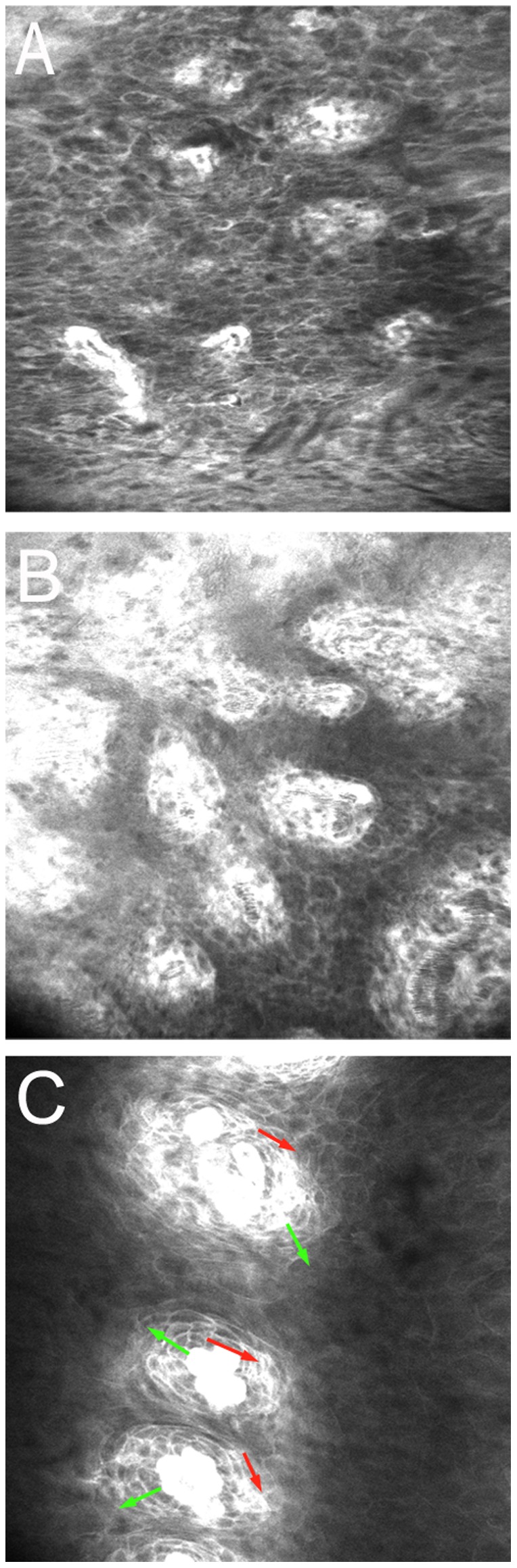
Confocal laser endomicroscopy (CLE) can provide in vivo subcellular resolution images of esophageal lesions. However, the learning curve in interpreting CLE images of precancerous or early-stage esophageal squamous cancer is unknown. The goal of this study is to evaluate the diagnostic accuracy and inter-observer agreement for differentiating esophageal lesions in CLE images among experienced and inexperienced observers and to assess the learning curve.
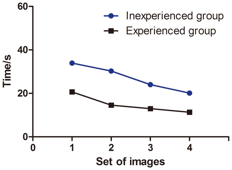
After a short training, 8 experienced and 14 inexperienced endoscopists evaluated in sequence 4 sets of high-quality CLE images. Their diagnoses were corrected and discussed after each set. For each image, the diagnostic results, confidence in diagnosis, quality and time to evaluate were recorded.
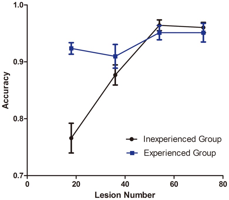
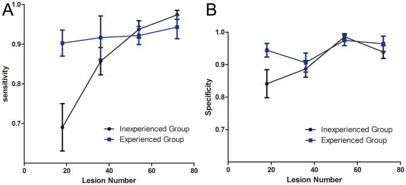
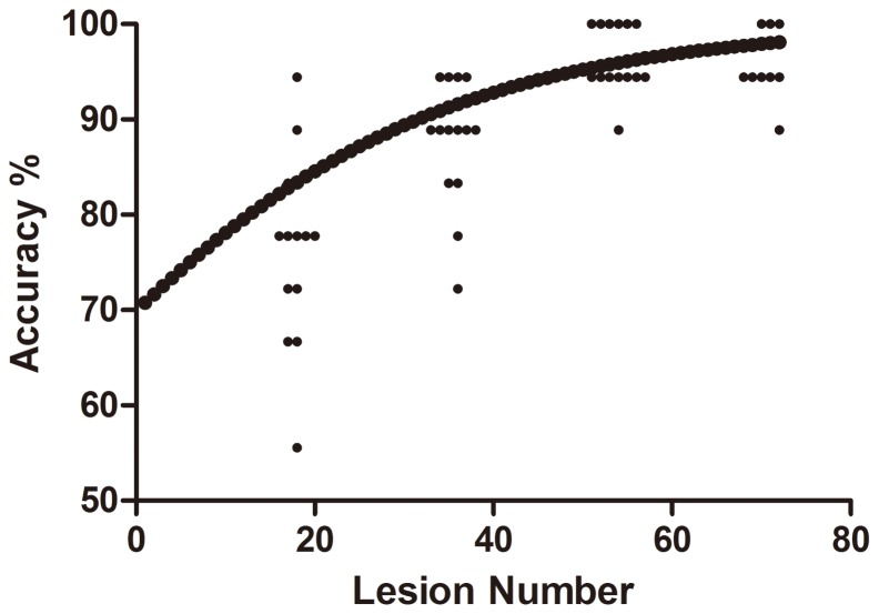
Overall, diagnostic accuracy was greater for the second, third, fourth set of images as compared with the initial set (odds ratio [OR] 2.01, 95% CI 1.22-3.31; 7.95, 3.74-16.87; and 6.45, 3.14-13.27), respectively, with no difference between the third and fourth sets in accuracy (p = 0.67). Previous experience affected the diagnostic accuracy only in the first set of images (OR 3.70, 1.87-7.29, p<0.001). Inter-observer agreement was higher for experienced than inexperienced endoscopists (0.732 vs. 0.666, p<0.01).
CLE is a promising technology that can be quickly learned after a short training period; previous experience is associated with diagnostic accuracy only at the initial stage of learning.
共聚焦激光内镜显微镜(CLE)能够提供食管病变的体内亚细胞分辨率图像。然而,解读癌前或早期食管鳞状癌CLE图像的学习曲线尚不清楚。本研究的目的是评估经验丰富和经验不足的观察者在CLE图像中鉴别食管病变的诊断准确性和观察者间一致性,并评估学习曲线。
经过简短培训后,8名经验丰富的内镜医师和14名经验不足的内镜医师依次评估4组高质量的CLE图像。每组图像评估后对他们的诊断进行校正和讨论。记录每张图像的诊断结果、诊断信心、图像质量和评估时间。
总体而言,与初始组相比,第二、第三、第四组图像的诊断准确性更高(优势比[OR]分别为2.01,95%置信区间1.22 - 3.31;7.95,3.74 - 16.87;以及6.45,3.14 - 13.27),第三组和第四组在准确性上无差异(p = 0.67)。既往经验仅在第一组图像中影响诊断准确性(OR 3.70,1.87 - 7.29,p<0.001)。经验丰富的内镜医师比经验不足的内镜医师观察者间一致性更高(0.732对0.666,p<0.01)。
CLE是一项有前景的技术,经过短时间培训后可快速掌握;既往经验仅在学习初期与诊断准确性相关。