Division of Gastroenterology, Long Island Jewish Medical Center, Zucker School of Medicine at Hofstra/Northwell, Northwell Health System, New Hyde Park, NY 11040, United States.
World J Gastroenterol. 2019 Jul 7;25(25):3108-3115. doi: 10.3748/wjg.v25.i25.3108.
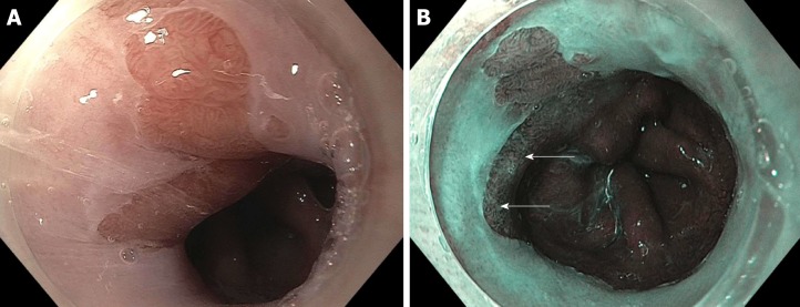
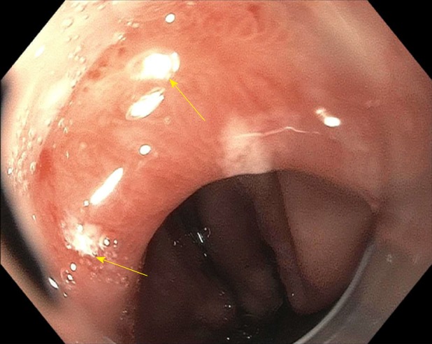
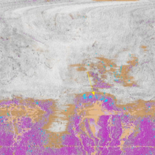
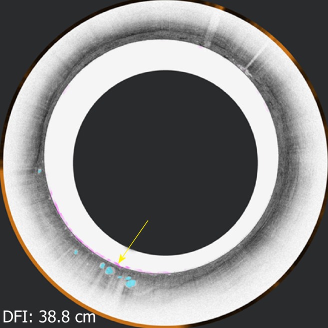
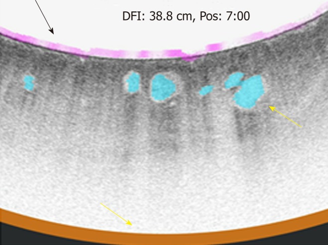
Esophageal cancer is on the rise. The known precursor lesion is Barrett's esophagus (BE). Patients with dysplasia are at higher risk of developing esophageal cancer. Currently the gold standard for surveillance endoscopy involves taking targeted biopsies of abnormal areas as well as random biopsies every 1-2 cm of the length of the Barrett's. Unfortunately studies have shown that this surveillance can miss dysplasia and cancer. Advanced imaging technologies have been developed that may help detect dysplasia in BE. This opinion review discusses advanced imaging in BE surveillance endoscopy and its utility in clinical practice.
食管癌发病率呈上升趋势。已知的癌前病变是巴雷特食管(BE)。有异型增生的患者发生食管癌的风险更高。目前,监测内镜的金标准包括对异常区域进行靶向活检,以及对 BE 长度每 1-2 厘米进行随机活检。遗憾的是,研究表明这种监测可能会遗漏异型增生和癌症。已经开发出了一些先进的成像技术,这些技术可能有助于检测 BE 中的异型增生。本综述讨论了 BE 监测内镜中的先进成像技术及其在临床实践中的应用。